
Page 526 - Feline diagnostic imaging
P. 526
538 30 Peritoneal Cavity
(b)
(a)
(c) (d)
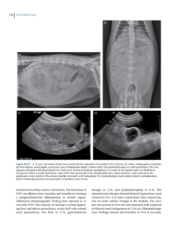
Figure 30.14 A 15-year-old mixed-breed male presented for evaluation of elevated liver enzymes. (a) Lateral radiographic projection.
(b) Ventrodorsal radiographic projection. Loss of abdominal detail is noted within the peritoneal space on both projections. The liver
appears enlarged, extending beyond the costal arch. Ventral and lateral spondylosis is present in the lumbar spine. (c) Abdominal
ultrasound shows a small hypoechoic mass within the spleen. (d) In the caudal abdomen, a more anechoic mass is found in the
peritoneal cavity cranial to the urinary bladder consistent with metastasis. On histopathologic examination, hepatic cystadenomas,
splenic hemangiosarcoma, and pancreatic amyloidosis were found.
mutation from feline enteric coronavirus. The two forms of changes in 2/16, and lymphadenopathy in 9/16. The
FIPV are effusive from vasculitis and noneffusive forming reported renal changes showed bilateral hyperechoic renal
a pyogranulomatosis inflammation in several organs. cortices in 3/16, 5/16 with a hypoechoic outer cortical rim,
Abdominal ultrasonographic finding were reported in 16 and one with nodular changes in the medulla. The renal
cats with FIPV. The majority of cats had a normal‐appear- size was normal in 9/16 cats and abnormal with unilateral
ing liver and spleen parenchyma, nearly half with normal or bilateral renal enlargement in 7/16 cats. Histopathologic
renal parenchyma, free fluid in 7/16, gastrointestinal renal findings showed abnormalities in 9/14 at necropsy.