
Page 529 - Feline diagnostic imaging
P. 529
30.4 Alternate Modalities 541
(a) (b)
(c)
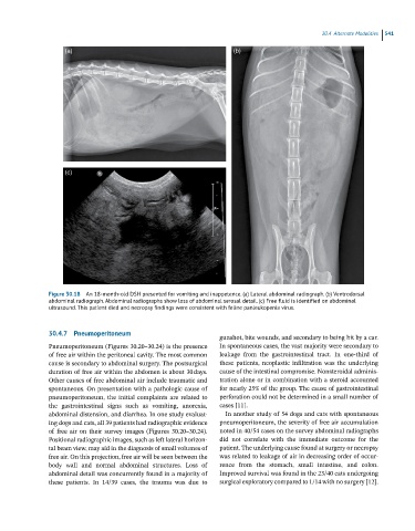
Figure 30.18 An 18-month-old DSH presented for vomiting and inappetence. (a) Lateral abdominal radiograph. (b) Ventrodorsal
abdominal radiograph. Abdominal radiographs show loss of abdominal serosal detail. (c) Free fluid is identified on abdominal
ultrasound. This patient died and necropsy findings were consistent with feline panleukopenia virus.
30.4.7 Pneumoperitoneum
gunshot, bite wounds, and secondary to being hit by a car.
Pneumoperitoneum (Figures 30.20–30.24) is the presence In spontaneous cases, the vast majority were secondary to
of free air within the peritoneal cavity. The most common leakage from the gastrointestinal tract. In one‐third of
cause is secondary to abdominal surgery. The postsurgical these patients, neoplastic infiltration was the underlying
duration of free air within the abdomen is about 30 days. cause of the intestinal compromise. Nonsteroidal adminis-
Other causes of free abdominal air include traumatic and tration alone or in combination with a steroid accounted
spontaneous. On presentation with a pathologic cause of for nearly 25% of the group. The cause of gastrointestinal
pneumoperitoneum, the initial complaints are related to perforation could not be determined in a small number of
the gastrointestinal signs such as vomiting, anorexia, cases [11].
abdominal distension, and diarrhea. In one study evaluat- In another study of 54 dogs and cats with spontaneous
ing dogs and cats, all 39 patients had radiographic evidence pneumoperitoneum, the severity of free air accumulation
of free air on their survey images (Figures 30.20–30.24). noted in 40/54 cases on the survey abdominal radiographs
Positional radiographic images, such as left lateral horizon- did not correlate with the immediate outcome for the
tal beam view, may aid in the diagnosis of small volumes of patient. The underlying cause found at surgery or necropsy
free air. On this projection, free air will be seen between the was related to leakage of air in decreasing order of occur-
body wall and normal abdominal structures. Loss of rence from the stomach, small intestine, and colon.
abdominal detail was concurrently found in a majority of Improved survival was found in the 23/40 cats undergoing
these patients. In 14/39 cases, the trauma was due to surgical exploratory compared to 1/14 with no surgery [12].