
Page 527 - Feline diagnostic imaging
P. 527
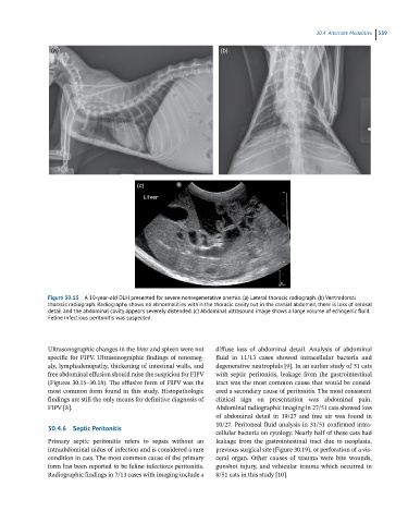
30.4 Alternate Modalities  539

                (a)                                                (b)


                                          (c)


               Figure 30.15  A 10-year-old DLH presented for severe nonregenerative anemia. (a) Lateral thoracic radiograph. (b) Ventrodorsal
               thoracic radiograph. Radiography shows no abnormalities within the thoracic cavity but in the cranial abdomen, there is loss of serosal
               detail and the abdominal cavity appears severely distended. (c) Abdominal ultrasound image shows a large volume of echogenic fluid.
               Feline infectious peritonitis was suspected.


               Ultrasonographic changes in the liver and spleen were not   diffuse  loss  of  abdominal  detail.  Analysis  of  abdominal
               specific for FIPV. Ultrasonographic findings of renomeg-  fluid  in  11/13  cases  showed  intracellular  bacteria  and
               aly, lymphadenopathy, thickening of intestinal walls, and   degenerative neutrophils [9]. In an earlier study of 51 cats
               free abdominal effusion should raise the suspicion for FIPV   with  septic  peritonitis,  leakage  from  the  gastrointestinal
               (Figures 30.15–30.18). The effusive form of FIPV was the   tract was the most common cause that would be consid-
               most common form found in this study. Histopathologic   ered a secondary cause of peritonitis. The most consistent
               findings are still the only means for definitive diagnosis of   clinical  sign  on  presentation  was  abdominal  pain.
               FIPV [8].                                          Abdominal radiographic imaging in 27/51 cats showed loss
                                                                  of  abdominal  detail  in  19/27  and  free  air  was  found  in
                                                                  10/27. Peritoneal fluid analysis in 31/51 confirmed intra-
               30.4.6  Septic Peritonitis
                                                                  cellular bacteria on cytology. Nearly half of these cats had
               Primary  septic  peritonitis  refers  to  sepsis  without  an   leakage  from  the  gastrointestinal  tract  due  to  neoplasia,
               intraabdominal nidus of infection and is considered a rare   previous surgical site (Figure 30.19), or perforation of a vis-
               condition in cats. The most common cause of the primary   ceral  organ.  Other  causes  of  trauma  were  bite  wounds,
               form has been reported to be feline infectious peritonitis.   gunshot injury, and vehicular trauma which occurred in
               Radiographic findings in 7/13 cases with imaging include a   8/51 cats in this study [10].