
Page 525 - Feline diagnostic imaging
P. 525
(a) (b)
(c)
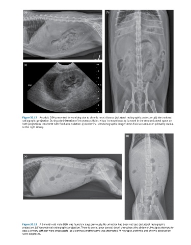
Figure 30.12 An adult DSH presented for vomiting due to chronic renal disease. (a) Lateral radiographic projection. (b) Ventrodorsal
radiographic projection. During administration of intravenous fluids, wispy increased opacity is noted in the retroperitoneal space on
both projections consistent with fluid accumulation. (c) Abdominal ultrasonographic image shows fluid accumulation primarily cranial
to the right kidney.
(b)
(a)
Figure 30.13 A 2-month-old male DSH was found six days previously. No urination had been noticed. (a) Lateral radiographic
projection. (b) Ventrodorsal radiographic projection. There is overall poor serosal detail throughout the abdomen. Multiple attempts to
pass a urinary catheter were unsuccessful so a perineal urethrostomy was attempted. At necropsy, urethritis and chronic obstruction
were diagnosed.