
Page 524 - Feline diagnostic imaging
P. 524
536 30 Peritoneal Cavity
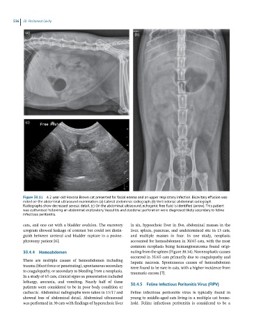
(a) (b)
(c)
Figure 30.11 A 2-year-old Havana Brown cat presented for facial edema and an upper respiratory infection. Bicavitary effusion was
noted on the abdominal ultrasound examination. (a) Lateral abdominal radiograph. (b) Ventrodorsal abdominal radiograph.
Radiographs show decreased serosal detail. (c) On the abdominal ultrasound, echogenic free fluid is identified (arrow). This patient
was euthanized following an abdominal exploratory. Vasculitis and duodenal perforation were diagnosed likely secondary to feline
infectious peritonitis.
cats, and one cat with a bladder avulsion. The excretory in six, hypoechoic liver in five, abdominal masses in the
urogram showed leakage of contrast but could not distin- liver, spleen, pancreas, and undetermined site in 13 cats,
guish between ureteral and bladder rupture in a postne- and multiple masses in four. In one study, neoplasia
phrotomy patient [6]. accounted for hemoabdomen in 30/65 cats, with the most
common neoplasia being hemangiosarcoma found origi-
30.4.4 Hemoabdomen nating from the spleen (Figure 30.14). Nonneoplastic causes
occurred in 35/65 cats primarily due to coagulopathy and
There are multiple causes of hemoabdomen including hepatic necrosis. Spontaneous causes of hemoabdomen
trauma (blunt force or penetrating), spontaneous secondary were found to be rare in cats, with a higher incidence from
to coagulopathy, or secondary to bleeding from a neoplasia. traumatic causes [7].
In a study of 65 cats, clinical signs on presentation included
lethargy, anorexia, and vomiting. Nearly half of these 30.4.5 Feline Infectious Peritonitis Virus (FIPV)
patients were considered to be in poor body condition or
cachectic. Abdominal radiographs were taken in 15/17 and Feline infectious peritonitis virus is typically found in
showed loss of abdominal detail. Abdominal ultrasound young to middle‐aged cats living in a multiple cat house-
was performed in 30 cats with findings of hyperechoic liver hold. Feline infectious peritonitis is considered to be a