
Page 10 - GP Fall 2022_Neat
P. 10
Treatment: All aphthous ulcers respond skin involvement. Any mucous membrane Treatment: This disease can only be treat-
well to steroids. Topical steroids are more sites can be affected, including oral, genital, ed palliatively. It cannot be cured as it is
effective on minor aphthous lesions. They ocular, and nasopharyngeal. Of significance, an autoimmune disease. Most patients with
can also be treated with ‘magic’ mouth 85% of patients present with oral lesions, limited disease can effectively be treated
rinses as well as topical mid-level steroids. often the initial manifestation of the disease, with corticosteroids (topical or systemic)
Minor aphthous ulcers will resolve on their and 65% present with ocular lesions. There with immunosuppressant medication as
own without scarring, however treatment is a special concern with ocular involvement needed. To maintain their oral health, it is
reduces the length of time and possibly the as eye lesions can lead to corneal desquama- recommended to gently brush with a soft
severity of the lesions. The biggest concern tion and conjunctival adhesions, known as toothbrush, use nonalcoholic based mouth
with patients on continuous doses of ste- symblepharon, which can result in scarring rinses and have frequent dental hygiene
roids is the possibility of adrenal suppres- and the potential for subsequent blindness. treatments. Remember that an eye referral
sion, therefore topical steroids should be As such, all patients with biopsy confirmed is always mandatory for patients with a di-
used whenever possible. mucous membrane pemphigoid must have a
referral for an eye exam. agnosis of mucous membrane pemphigoid.
Comparison of Herpes Simplex Virus vs.
Aphthous Ulcers Clinical: Mucous membrane pemphigoid is Pemphigus Vulgaris
considered to be a chronic and progressive
Secondary HSV1 lesions present with one disease. Patients often present with only oral Etiology: Pemphigus vulgaris is a muco-
of two clinical manifestations. They present lesions and the gingiva is the most common cutaneous, potentially fatal, autoimmune
either as herpes labialis, also referred to as site of occurrence followed by the buccal disease. There are many types of pemphi-
a cold sore or fever blister, or intraorally as mucosa. The gingiva presents as a desqua- gus, however, pemphigus vulgaris is the
ulcers on the attached alveolar mucosa, ke- mative gingivitis with a positive Nikolsky most common type and 80% of patients
ratinized gingiva and hard palate, referred sign, the ability to create a blister with gen- with pemphigus have pemphigus vulgaris.
to as herpes recurrans. In healthy people, in- tile rubbing. Scarring rarely develops with A biopsy is indicated to confirm the diag-
traoral herpetic lesions never present on the oral lesions. Confirmation of the diagnosis nosis of pemphigus vulgaris and blood tests
movable mucosa: cheeks, labial mucosa, is through a biopsy and direct immunohis- along with both indirect and direct immu-
buccal mucosa, soft palate, floor of mouth tochemistry looking for a positive linear nofluorescence can be used to confirm the
and underside of the tongue. Therefore, the epithelial immune deposit at the basement diagnosis. Indirect immunofluorescence
differential diagnosis of an unknown ulcer- membrane zone of the epithelium. The sero- identifies circulating pathogenic IgG an-
ation presenting on the intraoral attached logic detection of autoantibodies by indirect tibodies against desmoglien 3. In essence,
non-movable mucosa is by default auto- immunofluorescence is not always present the body is attacking the glue, desmoglien
matically reduced to either an iatrogenic or in patients with mucous membrane pemphi- 3, which holds the epithelial cells togeth-
traumatic ulceration or a secondary herpetic goid and the disease can be diagnosed on er. This produces a very fragile and easily
lesion. In the same fashion, intraoral ulcers biopsy alone (Figures 21, 22). ruptured intraepithelial blister. Before the
that occur on the movable mucosa will nev- advent of corticosteroids and immunosup-
er be herpetic in origin; they will either be pressant medications, pemphigus vulgaris
minor or traumatic ulcers. Although most was fatal in about 90% of all cases. Patients
clinicians are comfortable with diagnosing would die like burn victims die, from flu-
herpes labialis, the intraoral manifestations
of secondary herpetic lesions are often mis- id loss, dehydration, electrolyte imbalance
diagnosed. This is especially true in cases and subsequent kidney failure.
where blisters or vesicles were not present Clinical: Oral lesions are extremely com-
when the patient reported initially. It is im-
portant to remember that clinically this lack mon in early manifestations of pemphigus
of vesicles does not preclude the diagnosis vulgaris. Patients typically develop gingi-
of a secondary herpetic lesion. val lesions followed by lesions of the buc-
cal mucosa. The oral lesions run a chron-
Mucous Membrane Pemphigoid vs. ic course, causing fragile intraepithelial
Pemphigus Vulgaris blisters, which rupture easily resulting in
superficial erosions and ulcers. Gingival
Mucous Membrane Pemphigoid lesions present as a desquamative gingivi-
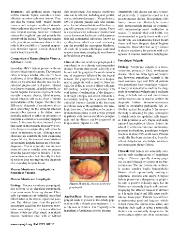
Figures 21 and 22. Mucous membrane tis with a positive Nikolsky sign, but the
Etiology: Mucous membrane pemphigoid, pemphigoid. blisters are extremely fragile and transient.
also referred to as cicatricial pemphigoid, Biopsying the affected mucosa is difficult
is an autoimmune blistering disease. Clini- as it is quite fragile and falls apart easily,
cally it presents as sub-epithelial tense fluid Age/Sex/Race: Mucous membrane pem- like wet tissue paper. Patients have difficul-
filled blisters at the dermal–epidermal junc- phigoid tends to present in the elderly pop- ty maintaining good oral hygiene, which,
tion. The blisters result from the patient’s ulation with a female predominance. It is in turn makes the lesions more active, and
autoantigen attacking the basement mem- rarely seen in children. It is frequently seen brushing their teeth or using a removable
brane zone antigen. It is a mucocutaneous in patients of Ashkenazi Jewish descent.
disease which can affect single or multiple denture can occasionally desquamate the
mucous membrane sites, with or without entire surface epithelium. Skin lesions tend
www.nysagd.org l Fall 2022 l GP 10