
Page 20 - GP Fall 2022_Neat
P. 20
Restoring Cervical Lesions with Microfilled Composite
Authors: Arthur R. Volker DDS, MSEd, FAGD, FACD and Aadel Soleymani, DDS
Introduction microfilled composite is radiolucency must be inserted at a steep angle, otherwise,
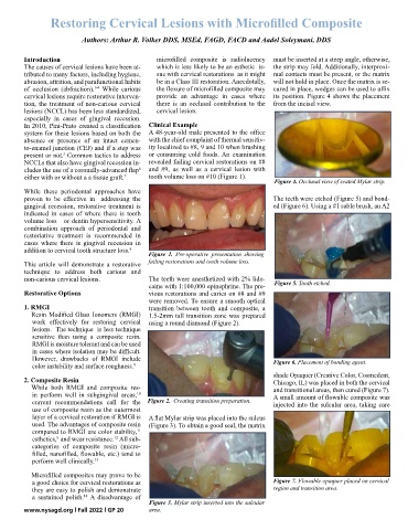
The causes of cervical lesions have been at- which is less likely to be an esthetic is- the strip may fold. Additionally, interproxi-
tributed to many factors, including hygiene, sue with cervical restorations as it might mal contacts must be present, or the matrix
abrasion, attrition, and parafunctional habits be in a Class III restoration. Anecdotally, will not hold in place. Once the matrix is se-
of occlusion (abfraction). While carious the flexure of microfilled composite may cured in place, wedges can be used to affix
1-4
cervical lesions require restorative interven- provide an advantage in cases where its position. Figure 4 shows the placement
tion, the treatment of non-carious cervical there is an occlusal contribution to the from the incisal view.
lesions (NCCL) has been less standardized, cervical lesion.
especially in cases of gingival recession.
In 2010, Pini-Prato created a classification Clinical Example
system for these lesions based on both the A 48-year-old male presented to the office
absence or presence of an intact cemen- with the chief complaint of thermal sensitiv-
to-enamel junction (CEJ) and if a step was ity localized to #8, 9 and 10 when brushing
present or not. Common tactics to address or consuming cold foods. An examination
5
NCCLs that also have gingival recession in- revealed failing cervical restorations on #8
cludes the use of a coronally-advanced flap and #9, as well as a cervical lesion with
6
either with or without a a tissue graft. 7 tooth volume loss on #10 (Figure 1).
Figure 4. Occlusal view of seated Mylar strip.
While these periodontal approaches have
proven to be effective in addressing the The teeth were etched (Figure 5) and bond-
gingival recession, restorative treatment is ed (Figure 6). Using a #1 sable brush, an A2
indicated in cases of where there is tooth
volume loss or dentin hypersensitivity. A
combination approach of periodontal and
restoriative treatment is recommended in
cases where there is gingival recession in
addition to cervical tooth structure loss. 8
Figure 1. Pre-operative presentation showing
This article will demonstrate a restorative failing restorations and tooth volume loss.
technique to address both carious and
non-carious cervical lesions. The teeth were anesthetized with 2% lido-
caine with 1:100,000 epinephrine. The pre- Figure 5. Tooth etched.
Restorative Options vious restorations and caries on #8 and #9
were removed. To ensure a smooth optical
1. RMGI transition between tooth and composite, a
Resin Modified Glass Ionomers (RMGI) 1.5-2mm tall transition zone was prepared
work effectively for restoring cervical using a round diamond (Figure 2).
lesions. The technique is less technique
sensitive than using a composite resin.
RMGI is moisture tolerant and can be used
in cases where isolation may be difficult.
However, drawbacks of RMGI include Figure 6. Placement of bonding agent.
color instability and surface roughness. 9
shade Opaquer (Creative Color, Cosmedent,
2. Composite Resin Chicago, IL) was placed in both the cervical
While both RMGI and composite res- and transitional areas, then cured (Figure 7).
in perform well in subgingival areas, A small amount of flowable composite was
10
current recommendations call for the Figure 2. Creating transition preparation. injected into the sulcular area, taking care
use of composite resin as the outermost
layer of a cervical restoration if RMGI is A flat Mylar strip was placed into the sulcus
used. The advantages of composite resin (Figure 3). To obtain a good seal, the matrix
compared to RMGI are color stability,
11
esthetics, and wear resistance. All sub-
12
8
categories of composite resin (micro-
filled, nanofilled, flowable, etc.) tend to
perform well clinically. 13
Microfilled composites may prove to be
a good choice for cervical restorations as Figure 7. Flowable opaquer placed on cervical
they are easy to polish and demonstrate region and transition area.
a sustained polish. A disadvantage of
14
Figure 3. Mylar strip inserted into the sulcular
www.nysagd.org l Fall 2022 l GP 20 area.