Page 175 - Essential Haematology
P. 175

Chapter 11  Haematological malignancy: aetiology and genetics  /  161


                                                                                              p15.2


                                                                                              q31
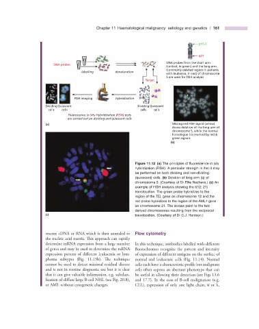
                                                                               DNA probes from the short arm
                          DNA probes
                                                                               (control, in green) and the long arm
                                                                               (commonly deleted region in patients
                                       labelling       denaturation
                                                                               with leukemia, in red) of chromosome
                                                                               5 are used for FISH analysis
                                                                     Target

                                    FISH imaging       hybridization

                      Dividing Quiescent                          Dividing Quiescent
                        cells  cells                               cells  cells
                                 Fluorescence In Situ Hybridization (FISH) tests
                                 are carried out on dividing and quiescent cells
                      (a)                                                         Missing red FISH signal (arrow)
                                                                                  shows deletion of the long arm of
                                                                                  chromosome 5, while the normal
                                                                                  homologue 5 is marked by red &
                                                                                  green signals
                                                                                 (b)


                                                                          Figure 11.12   (a)  The principles of fl uorescence  in situ


                                                                hybridization (FISH). A particular strength is that it may
                                                                be performed on both dividing and non - dividing
                                                                (quiescent) cells.  (b)  Deletion of long arm (q) of
                                                                chromosome 5. (Courtesy of Dr Ellie Nacheva.)  (c)  An
                                                                example of FISH analysis showing the t(12; 21)
                                                                translocation. The green probe hybridizes to the
                                                                region of the  TEL  gene on chromosome 12 and the
                                                                red probe hybridizes to the region of the  AML1  gene
                                                                on chromosome 21. The arrows point to the two
                                                                derived chromosomes resulting from the reciprocal
                      (c)                                       translocation.  (Courtesy of Dr C.J. Harrison.)


                      rescent cDNA or RNA which is then annealed to   Flow  c ytometry
                      the nucleic acid matrix. This approach can rapidly

                      determine mRNA expression from a large number   In this technique, antibodies labelled with diff erent
                      of genes and may be used to determine the mRNA  fl uorochromes recognize the pattern and intensity

                      expression pattern of different leukaemia or lym-  of expression of different antigens on the surface of

                      phoma subtypes (Fig.  11.13 b). Th e  technique  normal and leukaemic cells (Fig.  11.14 ). Normal
                      cannot be used to detect minimal residual disease  cells each have a characteristic profi le but malignant
                      and is not in routine diagnostic use but it is clear  cells often express an aberrant phenotype that can
                      that it can give valuable information, e.g. subclass-  be useful in allowing their detection (see Figs  13.6


                      fication of diffuse large B - cell NHL (see Fig.  20.8 ),  and  17.7   ). In the case of B - cell malignances (e.g.


                      or AML without cytogenetic changes.         CLL), expression of only one light chain,  κ  or  λ ,
   170   171   172   173   174   175   176   177   178   179   180