
Page 174 - Essential Haematology
P. 174
160 / Chapter 11 Haematological malignancy: aetiology and genetics
3' IgHC
5' 5'
C 5' IgHJ
c-MYC IgHV IgH J
V 5'
3' 8 8q- 3' 14 14q+ c-MYC
3'
t(8;14) (q24;q32)
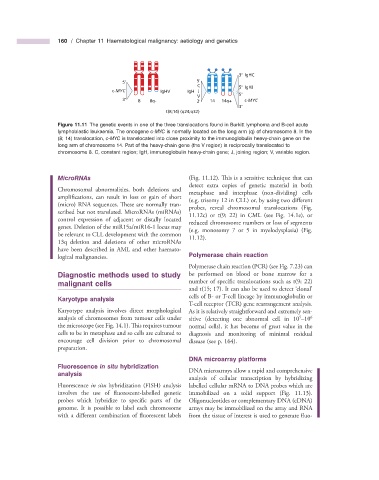
Figure 11.11 The genetic events in one of the three translocations found in Burkitt lymphoma and B - cell acute
lymphoblastic leukaemia. The oncogene c - MYC is normally located on the long arm (q) of chromosome 8. In the
(8; 14) translocation, c - MYC is translocated into close proximity to the immunoglobulin heavy - chain gene on the
long arm of chromosome 14. Part of the heavy - chain gene (the V region) is reciprocally translocated to
chromosome 8. C, constant region; IgH, immunoglobulin heavy - chain gene; J, joining region; V, variable region.
MicroRNAs (Fig. 11.12 ). This is a sensitive technique that can
detect extra copies of genetic material in both
Chromosomal abnormalities, both deletions and metaphase and interphase (non - dividing) cells
amplifications, can result in loss or gain of short (e.g. trisomy 12 in CLL) or, by using two diff erent
(micro) RNA sequences. These are normally tran- probes, reveal chromosomal translocations (Fig.
scribed but not translated. MicroRNAs (miRNAs) 11.12 c) or t(9; 22) in CML (see Fig. 14.1 e ), or
control expression of adjacent or distally located reduced chromosome numbers or loss of segments
genes. Deletion of the miR15a/miR16 - 1 locus may (e.g. monosomy 7 or 5 in myelodysplasia) (Fig.
be relevant to CLL development with the common 11.12 ).
13q deletion and deletions of other microRNAs
have been described in AML and other haemato-
logical malignancies. Polymerase c hain r eaction
Polymerase chain reaction (PCR) (see Fig. 7.23 ) can
Diagnostic m ethods u sed to s tudy be performed on blood or bone marrow for a
m alignant c ells number of specific translocations such as t(9; 22)
and t(15; 17). It can also be used to detect clonal ’
‘
cells of B - or T - cell lineage by immunoglobulin or
Karyotype a nalysis
T - cell receptor (TCR) gene rearrangement analysis.
Karyotype analysis involves direct morphological As it is relatively straightforward and extremely sen-
6
5
analysis of chromosomes from tumour cells under sitive (detecting one abnormal cell in 10 – 10
the microscope (see Fig. 14.1 ). This requires tumour normal cells), it has become of great value in the
cells to be in metaphase and so cells are cultured to diagnosis and monitoring of minimal residual
encourage cell division prior to chromosomal disease (see p. 164 ).
preparation.
DNA m icroarray p latforms
Fluorescence in s itu h ybridization
a nalysis DNA microarrays allow a rapid and comprehensive
analysis of cellular transcription by hybridizing
Fluorescence in situ hybridization (FISH) analysis labelled cellular mRNA to DNA probes which are
involves the use of fluorescent - labelled genetic immobilized on a solid support (Fig. 11.13 ).
probes which hybridize to specific parts of the Oligonucleotides or complementary DNA (cDNA)
genome. It is possible to label each chromosome arrays may be immobilized on the array and RNA
with a different combination of fl uorescent labels from the tissue of interest is used to generate fl uo-