
Page 170 - Essential Haematology
P. 170
156 / Chapter 11 Haematological malignancy: aetiology and genetics
and are called haploid . The chromosomes occur
in pairs and are numbered 1 – 22 in decreasing
size order; there are two sex chromosomes, XX
in females, XY in males. Karyotype is the term
used to describe the chromosomes derived from a
mitotic cell which have been set out in numerical
order (Fig. 11.6 ). A somatic cell with more or
less than 46 chromosomes is termed aneuploid ;
more than 46 is hyperdiploid , less than 46 hypodip-
loid ; 46 but with chromosome rearrangements,
pseudodiploid .
Each chromosome has two arms: the shorter
called ‘ p ’ , the longer called ‘ q ’ . These meet at the
centromere and the ends of the chromosomes are
called telomeres . On staining each arm divides into
regions numbered outwards from the centromere
and each region divides into bands (Fig. 11.7 ).
When a whole chromosome is lost or gained,
a − or + is put in front of the chromosome number.
If part of the chromosome is lost it is prefi xed with
del (for deletion). If there is extra material replacing
part of a chromosome the prefix add (for additional
material) is used. Chromosome translocations are
denoted by t, the chromosomes involved placed in
brackets with the lower numbered chromosome
fi rst. Th e prefix inv describes an inversion where
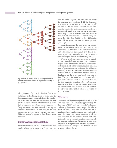
Figure 11.5 Multistep origin of a malignant tumour.
part of the chromosome has been inverted to run
Successive mutations lead to a growth advantage of
in the opposite direction. An isochromosome ,
one subclone.
denoted by i, describes a chromosome with identi-
cal chromosome arms at each end; for example,
i(17q) would consist of two copies of 17q joined at
the centromere.
lular pathways (Fig. 11.5 ). Another feature of
malignancy is clonal progression. In many cases the
Telomeres
disease develops new characteristics during its clini-
cal course and this may be accompanied by new Telomeres are repetitive sequences at the ends of
genetic changes. Selection of subclones may occur chromosomes. Th ey decrease by approximately 200
during treatment or reflect disease acceleration. base pairs of DNA with every round of replication.
Drug resistance can arise through a variety of When they decrease to a critical length, the cell exits
molecular mechanisms. In one example the cells from cell cycle. Germ cells and stem cells, which
express a protein that actively pumps a number of need to self - renew and maintain a high proliferative
different drugs to the outside of the cells (multidrug potential, contain the enzyme telomerase which can
resistance). add extensions to the telomeric repeats and com-
pensate for loss at replication and so enable the cells
to continue proliferation. Telomerase is also often
Chromosome n omenclature
expressed in malignant cells but this is probably a
The normal somatic cell has 46 chromosomes and consequence of the malignant transformation rather
is called diploid ; ova or sperm have 23 chromosomes than an initiating factor.