
Page 173 - Essential Haematology
P. 173
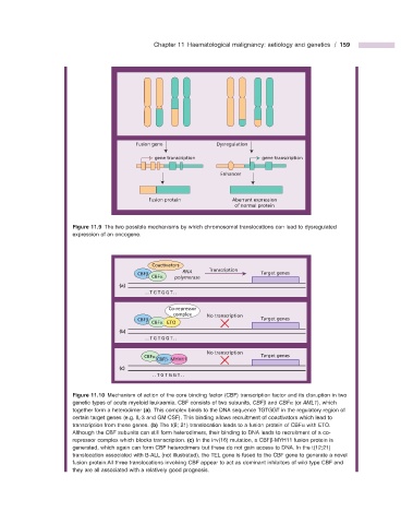
Chapter 11  Haematological malignancy: aetiology and genetics  /  159


                                         Fusion gene             Dysregulation

                                               gene transcription             gene transcription

                                                                  Enhancer


                                             Fusion protein          Aberrant expression
                                                                      of normal protein


                                Figure 11.9   The two possible mechanisms by which chromosomal translocations can lead to dysregulated

                      expression of an oncogene.


                                              Coactivators
                                          CBFβ         RNA     Transcription  Target genes
                                              CBFα  polymerase
                                    (a)
                                            ..TGTGGT..

                                                   Co-repressor
                                                    complex   No transcription
                                          CBFβ                                Target genes
                                              CBFα ETO
                                    (b)
                                            ..TGTGGT..
                                                              No transcription
                                           CBFα                               Target genes
                                               CBFβ- MYH11
                                    (c)
                                              ..TGTGGT..


                                Figure 11.10   Mechanism of action of the core binding factor (CBF) transcription factor and its disruption in two


                      genetic types of acute myeloid leukaemia. CBF consists of two subunits,  CBF β  and  CBF α  (or  AML1 ), which
                      together form a heterodimer  (a) . This complex binds to the DNA sequence TGTGGT in the regulatory region of
                      certain target genes (e.g. IL - 3 and GM - CSF). This binding allows recruitment of coactivators which lead to
                      transcription from these genes.  (b)  The t(8; 21) translocation leads to a fusion protein of CBF α  with ETO.
                      Although the CBF subunits can still form heterodimers, their binding to DNA leads to recruitment of a co -

                        repressor complex which blocks transcription.  (c)  In the inv(16) mutation, a CBF β - MYH11 fusion protein is
                      generated, which again can form CBF heterodimers but these do not gain access to DNA. In the t(12;21)
                      translocation associated with B - ALL (not illustrated), the TEL gene is fused to the CBF gene to generate a novel
                      fusion protein.All three translocations involving CBF appear to act as dominant inhibitors of wild type CBF and
                      they are all associated with a relatively good prognosis.