
Page 263 - Essential Haematology
P. 263
Chapter 19 Hodgkin lymphoma / 249
Table 19.2 Techniques for staging of lymphoma.
Laboratory Full blood count
ESR
Bone marrow aspirate and trephine (not routine)
Liver function
LDH
C - reactive protein
Radiology Chest X - ray
CT of thorax, abdomen, chest and pelvis
PET or PET/CT
MRI
Bone scan
CT, computed tomography; ESR, erythrocyte sedimentation rate; LDH, lactate
dehydrogenase; MRI, magnetic resonance imaging; PET, positron emission
tomography.
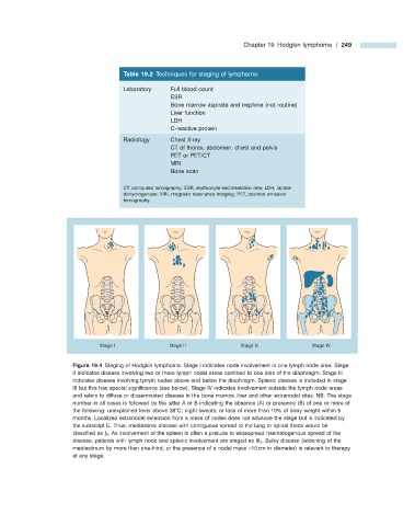
Stage I Stage II Stage III Stage IV
Figure 19.4 Staging of Hodgkin lymphoma. Stage I indicates node involvement in one lymph node area. Stage
II indicates disease involving two or more lymph nodal areas confi ned to one side of the diaphragm. Stage III
indicates disease involving lymph nodes above and below the diaphragm. Splenic disease is included in stage
III but this has special signifi cance (see below). Stage IV indicates involvement outside the lymph node areas
and refers to diffuse or disseminated disease in the bone marrow, liver and other extranodal sites. NB. The stage
number in all cases is followed by the letter A or B indicating the absence (A) or presence (B) of one or more of
the following: unexplained fever above 38 ° C; night sweats; or loss of more than 10% of body weight within 6
months. Localized extranodal extension from a mass of nodes does not advance the stage but is indicated by
the subscript E. Thus, mediastinal disease with contiguous spread to the lung or spinal theca would be
classifi ed as I E . As involvement of the spleen is often a prelude to widespread haematogenous spread of the
disease, patients with lymph node and splenic involvement are staged as III S . Bulky disease (widening of the
mediastinum by more than one - third, or the presence of a nodal mass > 10 cm in diameter) is relevant to therapy
at any stage.