
Page 260 - Essential Haematology
P. 260
246 / Chapter 19 Hodgkin lymphoma
Lymphomas are a group of diseases caused by malig-
nant lymphocytes that accumulate in lymph nodes
and cause the characteristic clinical features of lym-
phadenopathy. Occasionally, they may spill over
into blood ( ‘ leukaemic phase ’ ) or infi ltrate organs
outside the lymphoid tissue.
The major subdivision of lymphomas is into
Hodgkin lymphoma and non - Hodgkin lymphoma
and this is based on the histological presence
of Reed – Sternberg (RS) cells in Hodgkin
lymphoma.
History and p athogenesis
Thomas Hodgkin, curator of the anatomy museum
’
at Guy s Hospital in London, described the disease
in 1832. Dorothy Reed and Carl Sternberg were
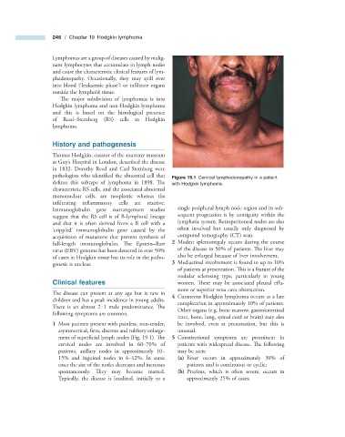
pathologists who identified the abnormal cell that Figure 19.1 Cervical lymphadenopathy in a patient
defines this subtype of lymphoma in 1898. Th e with Hodgkin lymphoma.
characteristic RS cells, and the associated abnormal
mononuclear cells, are neoplastic whereas the
infi ltrating inflammatory cells are reactive.
Immunoglobulin gene rearrangement studies single peripheral lymph node region and its sub-
suggest that the RS cell is of B - lymphoid lineage sequent progression is by contiguity within the
and that it is often derived from a B cell with a lymphatic system. Retroperitoneal nodes are also
‘ crippled ’ immunoglobulin gene caused by the often involved but usually only diagnosed by
acquisition of mutations that prevent synthesis of computed tomography (CT) scan.
full - length immunoglobulin. The Epstein – Barr 2 Modest splenomegaly occurs during the course
virus (EBV) genome has been detected in over 50% of the disease in 50% of patients. Th e liver may
of cases in Hodgkin tissue but its role in the patho- also be enlarged because of liver involvement.
genesis is unclear. 3 Mediastinal involvement is found in up to 10%
of patients at presentation. This is a feature of the
nodular sclerosing type, particularly in young
Clinical f eatures women. There may be associated pleural eff u-
sions or superior vena cava obstruction.
The disease can present at any age but is rare in
4 Cutaneous Hodgkin lymphoma occurs as a late
children and has a peak incidence in young adults.
complication in approximately 10% of patients.
There is an almost 2 : 1 male predominance. Th e
Other organs (e.g. bone marrow, gastrointestinal
following symptoms are common.
tract, bone, lung, spinal cord or brain) may also
1 Most patients present with painless, non - tender, be involved, even at presentation, but this is
asymmetrical, firm, discrete and rubbery enlarge- unusual.
ment of superfi cial lymph nodes (Fig. 19.1 ). Th e 5 Constitutional symptoms are prominent in
cervical nodes are involved in 60 – 70% of patients with widespread disease. Th e following
patients, axillary nodes in approximately 10 – may be seen:
15% and inguinal nodes in 6 – 12%. In some (a) Fever occurs in approximately 30% of
cases the size of the nodes decreases and increases patients and is continuous or cyclic;
spontaneously. They may become matted. (b) Pruritus, which is often severe, occurs in
Typically, the disease is localized, initially to a approximately 25% of cases;