
Page 262 - Essential Haematology
P. 262
248 / Chapter 19 Hodgkin lymphoma
(a) (b)
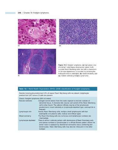
Figure 19.3 Hodgkin lymphoma: (a) high power view
of a lymph node biopsy showing two typical multi-
nucleate Reed – Sternberg cells, one with a characteris-
tic owl eye appearance, surrounded by lymphocytes,
histiocytes and an eosinophil; (b) mixed cellularity; and
(c) (c) nodular sclerosing Hodgkin lymphoma.
Table 19.1 World Health Organization (WHO) (2008) classifi cation of Hodgkin lymphoma.
Nodular lymphocyte - predominant (5% of cases) Reed – Sternberg cells are absent; lymphocyte
predominant (LP) tumour B cells are present
Classic Hodgkin lymphoma (95% of cases)
Nodular sclerosis Collagen bands extend from the node capsule to encircle nodules of
abnormal tissue. A characteristic lacunar cell variant of the Reed – Sternberg
cell is often found. The cellular infi ltrate may be of the lymphocyte -
predominant, mixed cellularity or lymphocyte - depleted type; eosinophilia is
frequent
Lymphocyte rich Scanty Reed – Sternberg cells; multiple small lymphocytes with few
eosinophils and plasma cells; nodular and diffuse types
Mixed cellularity The Reed – Sternberg cells are numerous and lymphocyte numbers are
intermediate
Lymphocyte depleted There is either a reticular pattern with dominance of Reed – Sternberg cells
and sparse numbers of lymphocytes or a diffuse fi brosis pattern where the
lymph node is replaced by disordered connective tissue containing few
lymphocytes. Reed – Sternberg cells may also be infrequent in this latter
subtype