
Page 261 - Essential Haematology
P. 261
Chapter 19 Hodgkin lymphoma / 247
(c) Alcohol - induced pain in the areas
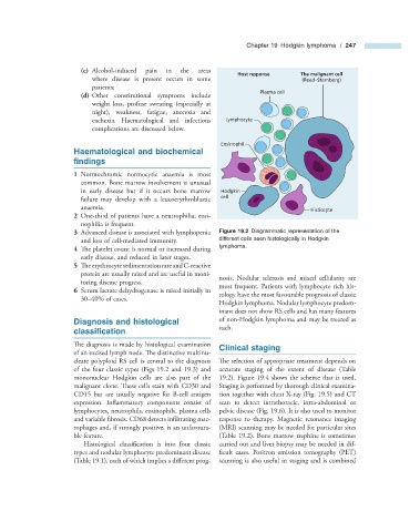
Host response The malignant cell
where disease is present occurs in some (Reed–Sternberg)
patients;
Plasma cell
(d) Other constitutional symptoms include
weight loss, profuse sweating (especially at
night), weakness, fatigue, anorexia and
cachexia. Haematological and infectious Lymphocyte
complications are discussed below.
Eosinophil
Haematological and b iochemical
fi ndings
1 Normochromic normocytic anaemia is most
common. Bone marrow involvement is unusual
in early disease but if it occurs bone marrow Hodgkin
failure may develop with a leucoerythroblastic cell
anaemia. Histiocyte
2 One - third of patients have a neutrophilia; eosi-
nophilia is frequent.
3 Advanced disease is associated with lymphopenia Figure 19.2 Diagrammatic representation of the
and loss of cell - mediated immunity. different cells seen histologically in Hodgkin
4 The platelet count is normal or increased during lymphoma.
early disease, and reduced in later stages.
5 The erythrocyte sedimentation rate and C - reactive
protein are usually raised and are useful in moni-
nosis. Nodular sclerosis and mixed cellularity are
toring disease progress.
most frequent. Patients with lymphocyte rich his-
6 Serum lactate dehydrogenase is raised initially in
tology have the most favourable prognosis of classic
30 – 40% of cases.
Hodgkin lymphoma. Nodular lymphocyte predom-
inant does not show RS cells and has many features
Diagnosis and h istological of non - Hodgkin lymphoma and may be treated as
such.
c lassifi cation
The diagnosis is made by histological examination Clinical s taging
of an excised lymph node. The distinctive multinu-
cleate polyploid RS cell is central to the diagnosis The selection of appropriate treatment depends on
of the four classic types (Figs 19.2 and 19.3 ) and accurate staging of the extent of disease (Table
mononuclear Hodgkin cells are also part of the 19.2 ). Figure 19.4 shows the scheme that is used.
malignant clone. These cells stain with CD30 and Staging is performed by thorough clinical examina-
CD15 but are usually negative for B - cell antigen tion together with chest X - ray (Fig. 19.5 ) and CT
expression. Inflammatory components consist of scan to detect intrathoracic, intra - abdominal or
lymphocytes, neutrophils, eosinophils, plasma cells pelvic disease (Fig. 19.6 ). It is also used to monitor
and variable fibrosis. CD68 detects infi ltrating mac- response to therapy. Magnetic resonance imaging
rophages and, if strongly positive, is an unfavoura- (MRI) scanning may be needed for particular sites
ble feature. (Table 19.2 ). Bone marrow trephine is sometimes
Histological classification is into four classic carried out and liver biopsy may be needed in dif-
types and nodular lymphocyte predominant disease ficult cases. Positron emission tomography (PET)
(Table 19.1 ), each of which implies a diff erent prog- scanning is also useful in staging and is combined