Page 329 - Essential Haematology
P. 329

Chapter 24  Platelets, blood coagulation and haemostasis  /  315


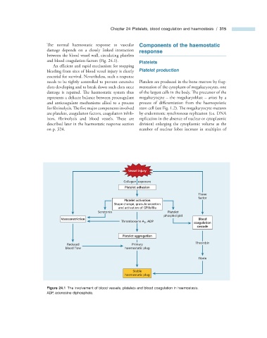
                       The normal haemostatic response to vascular       Components of the  h aemostatic
                      damage depends on a closely linked interaction     r esponse
                      between the blood vessel wall, circulating platelets
                      and blood coagulation factors (Fig.  24.1 ).           Platelets
                          An efficient and rapid mechanism for stopping


                      bleeding from sites of blood vessel injury is clearly       Platelet  p roduction
                      essential for survival. Nevertheless, such a response
                      needs to be tightly controlled to prevent extensive    Platelets are produced in the bone marrow by frag-
                      clots developing and to break down such clots once   mentation of the cytoplasm of megakaryocytes, one
                      damage is repaired. The haemostatic system thus   of the largest cells in the body. The precursor of the


                      represents a delicate balance between procoagulant   megakaryocyte  –  the megakaryoblast  –  arises by a
                      and anticoagulant mechanisms allied to a process   process of differentiation from the haemopoietic

                      for fi brinolysis. Th e five major components involved   stem cell (see Fig.  1.2 ). The megakaryocyte matures


                      are platelets, coagulation factors, coagulation inhib-  by endomitotic synchronous replication (i.e. DNA

                      itors, fibrinolysis and blood vessels. Th ese  are   replication in the absence of nuclear or cytoplasmic
                      described later in the haemostatic response section   division) enlarging the cytoplasmic volume as the
                      on    p. 324   .                          number of nuclear lobes increase in multiples of


                                                           Vessel injury

                                                         Collagen  exposure
                                                          Platelet adhesion
                                                                                           Tissue
                                                                                           factor
                                                          Platelet activation
                                                     Shape change, granule secretion
                                                       and activation of GPIIb/IIIa
                                              Serotonin                      Platelet
                                                                            phospholipid
                             Vasoconstriction                                              Blood
                                                                                         coagulation
                                                        Thromboxane A 2 , ADP
                                                                                           cascade
                                                         Platelet aggregation
                                                                                          Thrombin
                                Reduced                      Primary
                               blood flow                 haemostatic plug
                                                                                           Fibrin


                                                              Stable
                                                          haemostatic plug


                                Figure 24.1   The involvement of blood vessels, platelets and blood coagulation in haemostasis.


                      ADP, adenosine diphosphate.
   324   325   326   327   328   329   330   331   332   333   334