Page 331 - Essential Haematology
P. 331
(a)                                        (b)


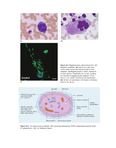
                                                                          Figure 24.3   Megakaryocytes:  (a)  immature form with
                                                                basophilic cytoplasm;  (b)  mature form with many
                                                                nuclear lobes and pronounced granulation of the
                                                                cytoplasm.  (c)  Megakaryocyte in culture, stained for
                                                                  α - tubulin (green). Proplatelets can be seen budding

                                                                from the tips of megakaryocyte cytoplasm.  (From
                        Control                                 Pecci A. et al. (2009)  Thrombosis and Haemostasis
                                                                  109 , 90 – 96, with permission; with thanks to Professor
                      (c)                                       Pecci for Fig.  24.3 c.)


                                                       Glycogen   Glycocalyx


                       Electron dense granule:
                                                                                 Lysosome
                       nucleotides (ADP),
                        2+
                       Ca , serotonin
                                                                                 Plasma membrane
                                                                                               Platelet
                                                                                               phospholipid
                       Specific α-granule:                                       Open canalicular
                       fibrinogen, factor V,                                     system
                       VWF, fibronectin,
                       heparin antagonist (PF 4),
                       PDGF, other proteins
                                                                                 Submembranous filaments
                                                                                 (platelet contractile protein)

                                                 Mitochondrion  Dense tubular system


                                Figure 24.4   The ultrastructure of platelets. ADP, adenosine diphosphate; PDGF, platelet - derived growth factor;


                      PF, platelet factor; VWF, von Willebrand factor.

