
Page 379 - Essential Haematology
P. 379
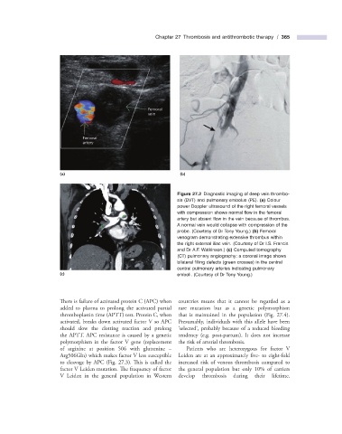
Chapter 27  Thrombosis and antithrombotic therapy  /  365


                      (a)                                        (b)


                                                                          Figure 27.2   Diagnostic imaging of deep vein thrombo-


                                                                sis (DVT) and pulmonary embolus (PE).  (a)  Colour
                                                                power Doppler ultrasound of the right femoral vessels
                                                                with compression shows normal fl ow in the femoral
                                                                artery but absent fl ow in the vein because of thrombus.
                                                                A normal vein would collapse with compression of the
                                                                probe. (Courtesy of Dr Tony Young.)  (b)  Femoral
                                                                venogram demonstrating extensive thrombus within
                                                                the right external iliac vein. (Courtesy of Dr I.S. Francis
                                                                and Dr A.F. Watkinson.)  (c)  Computed tomography
                                                                (CT) pulmonary angiography: a coronal image shows
                                                                bilateral fi lling defects (green crosses) in the central
                                                                central pulmonary arteries indicating pulmonary
                      (c)                                       emboli. (Courtesy of Dr Tony Young.)


                      There is failure of activated protein C (APC) when  countries means that it cannot be regarded as a

                      added to plasma to prolong the activated partial  rare mutation but as a genetic polymorphism
                      thromboplastin time (APTT) test. Protein C, when  that is maintained in the population (Fig.  27.4 ).
                      activated, breaks down activated factor V so APC  Presumably, individuals with this allele have been
                                                                ‘
                      should slow the clotting reaction and prolong    selected ’ , probably because of a reduced bleeding
                      the APTT. APC resistance is caused by a genetic  tendency (e.g. post - partum). It does not increase
                      polymorphism in the factor V gene (replacement  the risk of arterial thrombosis.
                      of arginine at position 506 with glutamine  –      Patients who are heterozygous for factor  V
                      Arg506Gln) which makes factor V less susceptible  Leiden are at an approximately five -  to eight - fold


                      to cleavage by APC (Fig.  27.3 ). This is called the  increased risk of venous thrombosis compared to

                      factor V Leiden mutation. The frequency of factor  the general population but only 10% of carriers
                      V Leiden in the general population in  Western  develop thrombosis during their lifetime.