
Page 42 - Essential Haematology
P. 42
28 / Chapter 2 Erythropoiesis and anaemia
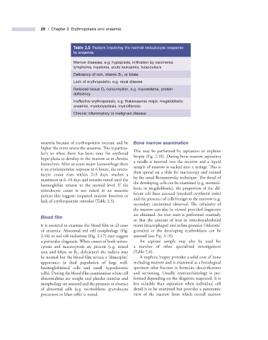
Table 2.5 Factors impairing the normal reticulocyte response
to anaemia.
Marrow diseases, e.g. hypoplasia, infi ltration by carcinoma,
lymphoma, myeloma, acute leukaemia, tuberculosis
Defi ciency of iron, vitamin B 12 or folate
Lack of erythropoietin, e.g. renal disease
Reduced tissue O 2 consumption, e.g. myxoedema, protein
defi ciency
Ineffective erythropoiesis, e.g. thalassaemia major, megaloblastic
anaemia, myelodysplasia, myelofi brosis
Chronic infl ammatory or malignant disease
anaemia because of erythropoietin increase and be Bone m arrow e xamination
higher the more severe the anaemia. This is particu- This may be performed by aspiration or trephine
larly so when there has been time for erythroid biopsy (Fig. 2.18 ). During bone marrow aspiration
hyperplasia to develop in the marrow as in chronic a needle is inserted into the marrow and a liquid
haemolysis. After an acute major haemorrhage there sample of marrow is sucked into a syringe. Th is is
is an erythropoietin response in 6 hours, the reticu- then spread on a slide for microscopy and stained
locyte count rises within 2 – 3 days, reaches a by the usual Romanowsky technique. The detail of
maximum in 6 – 10 days and remains raised until the the developing cells can be examined (e.g. normob-
haemoglobin returns to the normal level. If the lastic or megaloblastic), the proportion of the dif-
reticulocyte count is not raised in an anaemic ferent cell lines assessed (myeloid : erythroid ratio)
patient this suggests impaired marrow function or and the presence of cells foreign to the marrow (e.g.
lack of erythropoietin stimulus (Table 2.5 ).
secondary carcinoma) observed. The cellularity of
the marrow can also be viewed provided fragments
are obtained. An iron stain is performed routinely
Blood fi lm
so that the amount of iron in reticuloendothelial
It is essential to examine the blood film in all cases stores (macrophages) and as fine granules ( ‘ siderotic ’
of anaemia. Abnormal red cell morphology (Fig. granules) in the developing erythroblasts can be
2.16 ) or red cell inclusions (Fig. 2.17 ) may suggest assessed (see Fig. 3.10 ).
a particular diagnosis. When causes of both micro- An aspirate sample may also be used for
cytosis and macrocytosis are present (e.g. mixed a number of other specialized investigations
iron and folate or B 12 defi ciency) the indices may (Table 2.6 ).
be normal but the blood film reveals a ‘ dimorphic ’ A trephine biopsy provides a solid core of bone
appearance (a dual population of large well - including marrow and is examined as a histological
haemoglobinized cells and small hypochromic specimen after fixation in formalin, decalcifi cation
cells). During the blood film examination white cell and sectioning. Usually immunohistology is per-
abnormalities are sought and platelet number and formed depending on the diagnosis suspected. It is
morphology are assessed and the presence or absence less valuable than aspiration when individual cell
of abnormal cells (e.g. normoblasts, granulocyte detail is to be examined but provides a panoramic
precursors or blast cells) is noted. view of the marrow from which overall marrow