
Page 41 - Essential Haematology
P. 41
Chapter 2 Erythropoiesis and anaemia / 27
As well as suggesting the nature of the primary absence of other causes of macrocytosis (e.g. folate
defect, this approach may also indicate an underly- defi ciency).
ing abnormality before overt anaemia has
developed.
Other l aboratory fi ndings
In two common physiological situations the
mean corpuscular volume (MCV) may be outside Although the red cell indices will indicate the type
the normal adult range. In the newborn for a few of anaemia, further useful information can be
weeks the MCV is high but in infancy it is low (e.g. obtained from the initial blood sample.
70 fL at 1 year of age) and rises slowly throughout
childhood to the normal adult range. In normal
Leucocyte and p latelet c ounts
pregnancy there is a slight rise in MCV, even in the
‘
Measurement of these helps to distinguish pure ’
anaemia from ‘ pancytopenia ’ (subnormal levels of
red cells, neutrophils and platelets) which suggests
a more general marrow defect (e.g. caused by
marrow hypoplasia or infiltration) or general
destruction of cells (e.g. hypersplenism). In anae-
mias caused by haemolysis or haemorrhage, the
neutrophil and platelet counts are often raised; in
infections and leukaemias, the leucocyte count is
also often raised and there may be abnormal leuco-
cytes or neutrophil precursors present.
Reticulocyte c ount
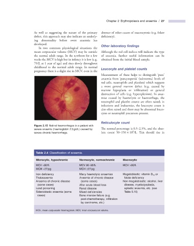
Figure 2.15 Retinal haemorrhages in a patient with
severe anaemia (haemoglobin 2.5 g/dL) caused by The normal percentage is 0.5 – 2.5%, and the abso-
9
severe chronic haemorrhage. lute count 50 – 150 × 10 /L. Th is should rise in
Table 2.4 Classifi cation of anaemia.
Microcytic, hypochromic Normocytic, normochromic Macrocytic
MCV < 80 fL MCV 80 – 95 fL MCV > 95 fL
MCH < 27 pg MCH ≥ 27 pg
Iron defi ciency Many haemolytic anaemias Megaloblastic: vitamin B 12 or
Thalassaemia Anaemia of chronic disease folate defi ciency
Anaemia of chronic disease (some cases) Non - megaloblastic: alcohol, liver
(some cases) After acute blood loss disease, myelodysplasia,
Lead poisoning Renal disease aplastic anaemia, etc. (see
Sideroblastic anaemia (some Mixed defi ciencies Table 5.10 )
cases) Bone marrow failure (e.g.
post - chemotherapy, infi ltration
by carcinoma, etc.)
MCH, mean corpuscular haemoglobin; MCV, mean corpuscular volume.