
Page 38 - Essential Haematology
P. 38
24 / Chapter 2 Erythropoiesis and anaemia
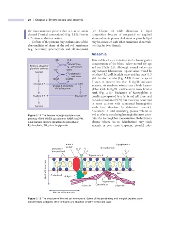
the transmembrane protein that acts as an anion (see Chapter 6 ) while alterations in lipid
channel ( ‘ vertical connections ’ ) (Fig. 2.12 ). Protein composition because of congenital or acquired
4.2 enhances this interaction. abnormalities in plasma cholesterol or phospholipid
Defects of the proteins may explain some of the may be associated with other membrane abnormali-
abnormalities of shape of the red cell membrane ties (e.g. in liver disease).
(e.g. hereditary spherocytosis and elliptocytosis)
Anaemia
H 2O 2 H 2O This is defined as a reduction in the haemoglobin
Glutathione concentration of the blood below normal for age
Embden-Meyerhof peroxidase
glycolytic pathway and sex (Table 2.3 ). Although normal values can
GSH GSSG
Glucose vary between laboratories, typical values would be
Glutathione less than 13.5 g/dL in adult males and less than 11.5
reductase
NADP NADPH g/dL in adult females (Fig. 2.13 ). From the age of
2 years to puberty, less than 11.0 g/dL indicates
Glucose-6-P 6-PG
Glucose-6-phosphate anaemia. As newborn infants have a high haemo-
dehydrogenase globin level, 14.0 g/dL is taken as the lower limit at
birth (Fig. 2.13 ). Reduction of haemoglobin is
Fructose-6-P Ribulose-5-P usually accompanied by a fall in red cell count and
packed cell volume (PCV) but these may be normal
in some patients with subnormal haemoglobin
Lactate
levels (and therefore by defi nition anaemic).
Alterations in total circulating plasma volume as
well as of total circulating haemoglobin mass deter-
Figure 2.11 The hexose monophosphate shunt
pathway. GSH, GSSG, glutathione; NADP, NADPH, mine the haemoglobin concentration. Reduction in
nicotinamide adenine dinucleotide phosphate; plasma volume (as in dehydration) may mask
P, phosphate; PG, phosphoglycerate. anaemia or even cause (apparent, pseudo) poly-
Band 3 Glycophorin B
protein Glycophorin C
Membrane
phospholipid
Glycophorin A
Vertical interaction Cholesterol 4.2 4.1 Actin
Ankyrin
4.1
Tropomyosin
Cytoskeleton
α Spectrin β Spectrin
Horizontal interaction
Figure 2.12 The structure of the red cell membrane. Some of the penetrating and integral proteins carry
carbohydrate antigens; other antigens are attached directly to the lipid layer.