
Page 151 - parasitology for medical and clinical laboratoryprofessionals
P. 151
Epidemiology of Nematodes, Cestodes, and Trematodes 131
Eggs carried to the heart produce arteriolitis (small
vessel inflammation) and fibrosis resulting in enlarge-
ment and failure of the right ventricle. As in other para-
sitic infections, the infiltration of tissues of the body serve
to stimulate production of large numbers of white blood
cells called eosinophils which are produced during seri-
ous allergic responses. Source: Centers for Disease Control and Prevention (CDC)
Pathological reactions are stimulated by the cercaria
upon contact with a potential host. The swimmer’s itch
is due to physical damage to the skin by proteases and
other toxic substances secreted by the invading cercaria.
A severe immunological reaction by the host may lead
to severe hypersensitivity (allergic) reactions to schis-
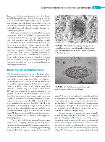
FIGURE 5-13 Schistosoma japonicum egg, and its
tosomal secretions and egg constituents. Embryonated
vestigial spine (arrow) taken from a liver tissue biopsy.
eggs cause collagenase-mediated damage to the vascular Eggs are smaller (55–65 μm by 70–100 μm) than those
endothelium. Host immune responses, both humoral of the other species
(blood plasma antibodies) and cell mediated, have been
shown to be of some protective value. IgE (immune
globulin increased in allergic reactions) and eosinophil-
mediated cytotoxicity have been hypothesized as a means
of killing the adult worm.
Diagnosis of Schistosomiasis Source: Centers for Disease Control and Prevention (CDC)
The diagnosis is based on a history of residence in or
travel to an endemic area, the development of swim-
mer’s itch, or other symptoms. The eggs of each of
the schistosomes are particularly characteristic and
are used to confirm diagnosis. S. hematobium eggs in
urine (55 to 65 by 110 to 170 μm) have an apical spine FIGURE 5-14 Schistosoma hematobium egg’s
or knob. S. mansoni eggs in feces (45 to 70 by 115 to posteriorly protruding, terminal spine
175 μm) have a spine on the side. S. japonicum eggs
(55 to 65 by 70 to 100 μm) are rounder than the other
two of this genus, and possess a vague spine on the side human’s feces or urine, and under optimal conditions
(Figure 5-13). In order to prevent and control schistoso- in a watery environment, the eggs hatch and release
miasis, contaminated water should be avoided. All fresh “miracidia,” which then penetrate a specific snail inter-
water bodies in endemic areas should be avoided, as the mediate host. Once inside the host, the S. haematobium
possibility of contamination is high. Control measures parasite passes through two developmental generations
include sanitary disposal of sewage that includes urine of sporocysts and are released by the snail into its envi-
and the destruction of species of snails capable or being ronment as “cercariae” (Figure 5-15).
hosts. Medications are available to control the infection Schistosoma mansoni parasites are found primarily
but there is no specific inoculation available to prevent in most areas of Africa and in tropical America, particu-
becoming infected. larly the West Indies. Mesenteric veins of the connec-
The egg of the Schistosoma haematobium trematode tions of the small intestine to the posterior abdominal
parasite has a unique posteriorly protruding, termi- membranes lining the cavity provide an anatomic site
nal spine unlike the vestigial remnant protruding from for these organisms to colonize. Eggs may be emit-
the lateral wall of the Schistosoma japonicum egg ted through either the bladder or through the intestine
(Figure 5-14). These eggs are eliminated in an infected (Figure 5-16). S. mansoni eggs are larger than those of