
Page 222 - parasitology for medical and clinical laboratoryprofessionals
P. 222
202 CHAPTER 9
LIFE CYCLE of—
Trichinella spiralis
Larva deposited
in mucosa
Adults in small intestine
Circulation
MAN
Larva released
in small intestine
Encysted larva in striated muscle
(diagnostic stage)
Adults in small intestine
Larva deposited
Larva released in mucosa
in small intestine
SWINE
OTHER CARNIVORES Circulation
Source: Centers for Disease Control and Prevention (CDC)
Ingested Encysted larva in
striated muscle
(diagnostic stage)
Encysted larva in
striated muscle
(infective stage)
MEAT
(PORK, etc.)
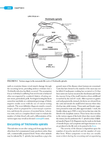
FIGURE 9-1 Various stages in the nematode life cycle of Trichinella spiralis
such as creatinine kinase reveal muscle damage through general cause of the disease where humans are concerned.
the encysting larvae, providing indirect evidence that a Cysts that have formed in the muscles of the meat may not
Trichinella infection has likely occurred. The assumption be killed if inadequate cooking has occurred or if it has
that an individual is suffering from trichinosis is bolstered been eaten raw. Larvae encyst in the duodenum and invade
when accompanied by a patient’s history of eating cer- the mucous lining of the small intestine where adulthood
tain meats, particularly pork. Serological testing has been is reached by the end of 1 week. After exposure to gastric
somewhat unreliable as a substantial percentage of falsely acid and pepsin in the stomach, the larvae are released from
negative results occur with the use of current testing the cysts and invade the small bowel mucosa where they
methods. The most definitive diagnosis entails a muscle develop into adult worms. These male and female organ-
biopsy, which is prepared for a microscopic examina- isms proceed to mate and, after approximately 1 week, the
tion, and shows the presence of encysted larvae. Labora- females release up to 1500 or more larvae that may enter the
tory tests, such as a complete blood count to evaluate the blood or the lymphatic system. These larvae then circulate
number of white blood cells and a differentiation of the to the various organs of the body where they encyst within
various types may reveal an elevated eosinophil count. the tissues, but the preference for T. spiralis is that of skele-
tal muscle (Figure 9-2). Diagnosis may be made on the basis
Encysting of Trichinella spiralis of muscle biopsy and symptoms and signs because no ova
are produced in the life cycle to be passed in the feces.
This infection occurs after eating pork from pigs that have The severity of the disease is dependent upon the
often been fed contaminated waste products rather than number of muscles involved and the number of cysts
safe, commercially prepared food. Some other animals that form. When symptoms occur they are usually
may be infected by T. spiralis, but meat from a pig is the most evident during the encysting and encapsulating