
Page 228 - parasitology for medical and clinical laboratoryprofessionals
P. 228
208 CHAPTER 9
6
SAFER•HEALTHIER•PEOPLE™
1
Scolex attaches http://www.dpd.cdc.gov/dpdx
to intestine
4
Adult in small intestine
5
2
Protoscolex
from cyst
Ingestion of cysts
(in organs) Definitive Host i 2
(dogs & other canidae) 4
4
Intermediate Host Embryonated 4
(sheep, goats, swine, etc.) Ingestion of eggs
(in feces) egg in feces 4
4
d 4 Source: Centers for Disease Control and Prevention (CDC)
3 3
i = Infective Stage
Oncosphere hatches; d = Diagnostic Stage
Hydatid cyst in liver, lungs, etc. penetrates intestinal wall
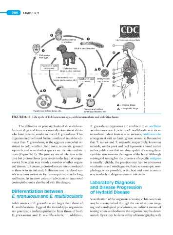
FIGURE 9-11 Life cycle of Echincococcus spp., with intermediate and definitive hosts
The definitive or primary hosts of E. multilocu- E. granulosus organisms are confined to an acellular
laris are dogs and foxes occasionally domesticated cats membranous vesicle, whereas E. multilocularis in its in-
who hunt rodents, similar to that of E. granulosus. This termediate rodent hosts is of an invasive, multivesicular
organism may be found farther north and in colder cli- arrangement with no limiting layer around it. Remember
mates than E. granulosus, as the eggs are somewhat re- that T. solium and T. saginata, respectively, known as
sistant to cold weather. Field mice, muskrats, ground taeniids, are the pork and beef tapeworms found earlier
squirrels, and several other species are the intermediate in this publication that are also capable of causing these
hosts (Figure 9-11). The primary site of infection is the cyst-like structures in the organs of the body. Although
liver but protoscoleces (precursors to the head of a tape- serological testing for the presence of specific antigens
worm) from cysts may invade a number of other organs is usually reliable, the practice may lead to erroneous
and tissues. In humans, protoscoleces are rarely produced conclusions and misdiagnosis. Basic microscopic mor-
in those who are infected. Infiltration into the blood ves- phology, when possible, is the best and most accurate
sels may cause metastatic formations primarily in the lung way in which to diagnose current infections.
and brain. As in most parasitic infections an increased
eosinophil count is also found with this disease. Laboratory Diagnosis
and Disease Progression
Differentiation between of Hydatid Disease
E. granulosus and E. multilocularis
Visualization of the organisms causing echinococcosis
Adult worms of E. granulosus are larger than those of may be accomplished through the use of various imag-
E. multilocularis. Eggs of the taeniid-type organisms ing and serological procedures, an indirect means of
are practically indistinguishable from those of both testing where antibodies to the organism may be deter-
E. granulosus and E. multilocularis. In addition, mined. Cysts may be detected by ultrasonography, with