
Page 270 - Atlas of Histology with Functional Correlations
P. 270
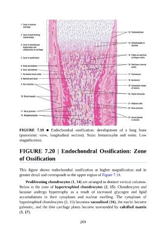
FIGURE  7.19  ■  Endochondral  ossification:  development  of  a  long  bone
               (panoramic  view,  longitudinal  section).  Stain:  hematoxylin  and  eosin.  Low
               magnification.


               FIGURE  7.20  |  Endochondral  Ossification:  Zone

               of Ossification


               This  figure  shows  endochondral  ossification  at  higher  magnification  and  in

               greater detail and corresponds to the upper region of Figure 7.19.

                   Proliferating chondrocytes (1, 14) are arranged in distinct vertical columns.
               Below is the zone of hypertrophied chondrocytes (2, 15). Chondrocytes and
               lacunae  undergo  hypertrophy  as  a  result  of  increased  glycogen  and  lipid

               accumulations  in  their  cytoplasm  and  nuclear  swelling.  The  cytoplasm  of
               hypertrophied chondrocytes (2, 15) becomes vacuolized (16), the nuclei become
               pyknotic, and the thin cartilage plates become surrounded by calcified matrix
               (5, 17).


                                                          269