
Page 275 - Atlas of Histology with Functional Correlations
P. 275
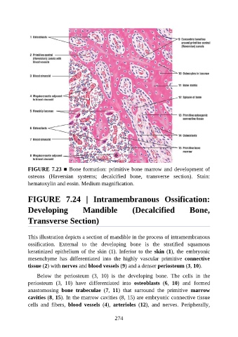
FIGURE 7.23 ■ Bone formation: primitive bone marrow and development of
               osteons  (Haversian  systems;  decalcified  bone,  transverse  section).  Stain:

               hematoxylin and eosin. Medium magnification.


               FIGURE  7.24  |  Intramembranous  Ossification:


               Developing                 Mandible                 (Decalcified                 Bone,

               Transverse Section)


               This illustration depicts a section of mandible in the process of intramembranous

               ossification.  External  to  the  developing  bone  is  the  stratified  squamous
               keratinized  epithelium  of  the  skin  (1).  Inferior  to  the  skin (1),  the  embryonic
               mesenchyme  has  differentiated  into  the  highly  vascular  primitive  connective
               tissue (2) with nerves and blood vessels (9) and a denser periosteum (3, 10).


                   Below  the  periosteum  (3,  10)  is  the  developing  bone.  The  cells  in  the
               periosteum  (3,  10)  have  differentiated  into  osteoblasts  (6,  10)  and  formed
               anastomosing  bone  trabeculae  (7,  11)  that  surround  the  primitive  marrow
               cavities (8, 15). In the marrow cavities (8, 15) are embryonic connective tissue
               cells  and  fibers,  blood  vessels  (4),  arterioles  (12),  and  nerves.  Peripherally,


                                                          274