
Page 272 - Atlas of Histology with Functional Correlations
P. 272
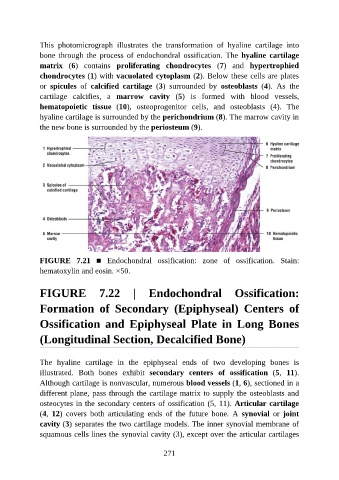
This  photomicrograph  illustrates  the  transformation  of  hyaline  cartilage  into

               bone  through  the  process  of  endochondral  ossification.  The  hyaline  cartilage
               matrix  (6)  contains  proliferating  chondrocytes  (7)  and  hypertrophied
               chondrocytes (1) with vacuolated cytoplasm (2). Below these cells are plates
               or  spicules  of  calcified  cartilage  (3)  surrounded  by  osteoblasts  (4).  As  the
               cartilage  calcifies,  a  marrow  cavity  (5)  is  formed  with  blood  vessels,

               hematopoietic  tissue  (10),  osteoprogenitor  cells,  and  osteoblasts  (4).  The
               hyaline cartilage is surrounded by the perichondrium (8). The marrow cavity in
               the new bone is surrounded by the periosteum (9).


               FIGURE  7.21  ■  Endochondral  ossification:  zone  of  ossification.  Stain:

               hematoxylin and eosin. ×50.


               FIGURE  7.22  |  Endochondral  Ossification:


               Formation  of  Secondary  (Epiphyseal)  Centers  of

               Ossification  and  Epiphyseal  Plate  in  Long  Bones


               (Longitudinal Section, Decalcified Bone)


               The  hyaline  cartilage  in  the  epiphyseal  ends  of  two  developing  bones  is
               illustrated.  Both  bones  exhibit  secondary  centers  of  ossification  (5,  11).
               Although cartilage is nonvascular, numerous blood vessels (1, 6), sectioned in a

               different plane, pass through the cartilage matrix to supply the osteoblasts and
               osteocytes in the secondary centers of ossification (5, 11). Articular cartilage
               (4,  12)  covers  both  articulating  ends  of  the  future  bone.  A  synovial  or  joint
               cavity (3) separates the two cartilage models. The inner synovial membrane of
               squamous cells lines the synovial cavity (3), except over the articular cartilages


                                                          271