
Page 274 - Atlas of Histology with Functional Correlations
P. 274
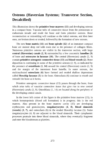
Osteons  (Haversian  Systems;  Transverse  Section,

               Decalcified)


               This illustration shows the primitive bone marrow (15) and developing osteons
               in a compact bone. Vascular tufts of connective tissue from the periosteum or

               endosteum  invade  and  erode  the  bone  and  form  primitive  osteons.  Bone
               reconstruction or remodeling will continue as the initial osteons, and then later
               ones, are broken down or eroded, followed by the formation of new osteons.

                   The new bone matrix (11) and bone spicule (12) of an immature compact
               bone  are  stained  deep  red  with  eosin  due  to  the  presence  of  collagen  fibers.

               Numerous  primitive  osteons  are  visible  in  the  transverse  section,  with  large
               central (Haversian) canals (2, 9) surrounded by a few concentric lamellae (9)
               of bone and osteocytes in lacunae (10). The central (Haversian) canals (2, 9)

               contain primitive osteogenic connective tissue (13) and blood vessels (2). Bone
               deposition is continuing in some of the primitive osteons (2, 9), as indicated by
               the presence of osteoblasts (1, 14) around the central (Haversian) canals (2, 9)
               and  the  margin  of  the  innermost  bone  lamella.  In  some  osteons,  the
               multinucleated  osteoclasts  (6)  have  formed  and  eroded  shallow  depressions

               called Howship lacunae (5) in the bone. Osteoclasts (6) continue to resorb and
               remodel the bone as it forms.

                   Primitive  osteogenic  connective  tissue  (13)  passes  through  the  bone,  from
               which  arise  tufts  of  vascular  connective  tissue  that  give  rise  to  new  central

               (Haversian) canals (2, 9). Osteoblasts (1, 14) are located along the periphery of
               the developing central canals.

                   In the lower left corner of the figure is the primitive bone marrow (15), in
               which  hematopoiesis  (blood  cell  formation)  is  in  progress;  this  is  the  red

               marrow.  Also  present  in  the  bone  marrow  cavity  (15)  are  developing
               erythrocytes  and  granulocytes,  megakaryocytes  (4,  8),  blood  sinusoids
               (vessels) (3, 7),  and  osteoclasts  (6)  in  the  eroded  Howship  lacunae  (5).  Some
               megakaryocytes  (4,  8)  are  adjacent  to  the  blood  sinusoids.  Their  cytoplasmic

               processes protrude into these blood sinusoids, where they eventually fragment
               and enter the bloodstream as platelets.


                                                          273