
Page 277 - Atlas of Histology with Functional Correlations
P. 277
(3) are the multinuclear osteoclasts (8) that remodel the developing bone. A
primitive marrow cavity (4) with blood vessels (9), blood cells (9), and
hematopoietic tissue is located between the formed bony trabeculae (3).
FIGURE 7.25 ■ Intramembranous ossification: developing skull bone
(decalcified bone; transverse section). Stain: Mallory-Azan. ×64.
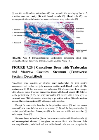
FIGURE 7.26 | Cancellous Bone with Trabeculae
and Marrow Cavities: Sternum (Transverse
Section, Decalcified)
Cancellous bone consists of slender bony trabeculae (5) that ramify,
anastomose, and enclose irregular marrow cavities with blood vessels (4). The
periosteum (2, 7) that surrounds the trabeculae (5) of cancellous bone merges
with adjacent dense irregular connective tissue with blood vessels (1). Inferior
to the periosteum (2, 7), the bony trabeculae (5) merge with a thin layer of
compact bone (9) that contains a forming or primitive osteon (6) and a mature
osteon (Haversian system) (8) with concentric lamellae.
Except for concentric lamellae in the primitive osteon (6) and the mature
osteon (8), the bone inferior to the periosteum (2, 7) and the bony trabeculae (5)
exhibit parallel lamellae. Osteocytes (3) in lacunae are visible in trabeculae (5)
and compact bone (9).
Between bony trabeculae (5) are the marrow cavities with blood vessels (4)
and hematopoietic tissue (11) that gives rise to new blood cells. Because of the
low magnification, individual red and white blood cells are not recognizable.
276