
Page 280 - Atlas of Histology with Functional Correlations
P. 280
both calcium and phosphate. These effects lower the circulating calcium
levels in the body. The actions of both thyroid and parathyroid glands and
their hormones are discussed in more detail in Chapter 19.
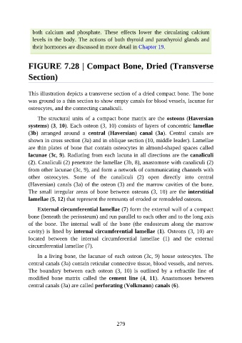
FIGURE 7.28 | Compact Bone, Dried (Transverse
Section)
This illustration depicts a transverse section of a dried compact bone. The bone
was ground to a thin section to show empty canals for blood vessels, lacunae for
osteocytes, and the connecting canaliculi.
The structural units of a compact bone matrix are the osteons (Haversian
systems) (3, 10). Each osteon (3, 10) consists of layers of concentric lamellae
(3b) arranged around a central (Haversian) canal (3a). Central canals are
shown in cross section (3a) and in oblique section (10, middle leader). Lamellae
are thin plates of bone that contain osteocytes in almond-shaped spaces called
lacunae (3c, 9). Radiating from each lacuna in all directions are the canaliculi
(2). Canaliculi (2) penetrate the lamellae (3b, 8), anastomose with canaliculi (2)
from other lacunae (3c, 9), and form a network of communicating channels with
other osteocytes. Some of the canaliculi (2) open directly into central
(Haversian) canals (3a) of the osteon (3) and the marrow cavities of the bone.
The small irregular areas of bone between osteons (3, 10) are the interstitial
lamellae (5, 12) that represent the remnants of eroded or remodeled osteons.
External circumferential lamellae (7) form the external wall of a compact
bone (beneath the periosteum) and run parallel to each other and to the long axis
of the bone. The internal wall of the bone (the endosteum along the marrow
cavity) is lined by internal circumferential lamellae (1). Osteons (3, 10) are
located between the internal circumferential lamellae (1) and the external
circumferential lamellae (7).
In a living bone, the lacunae of each osteon (3c, 9) house osteocytes. The
central canals (3a) contain reticular connective tissue, blood vessels, and nerves.
The boundary between each osteon (3, 10) is outlined by a refractile line of
modified bone matrix called the cement line (4, 11). Anastomoses between
central canals (3a) are called perforating (Volkmann) canals (6).
279