
Page 276 - Atlas of Histology with Functional Correlations
P. 276
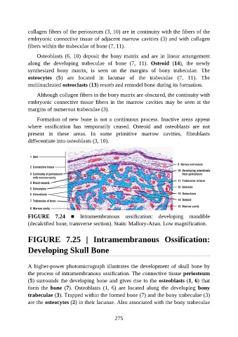
collagen fibers of the periosteum (3, 10) are in continuity with the fibers of the

               embryonic connective tissue of adjacent marrow cavities (3) and with collagen
               fibers within the trabeculae of bone (7, 11).

                   Osteoblasts  (6,  10)  deposit  the  bony  matrix  and  are  in  linear  arrangement
               along  the  developing  trabeculae  of  bone  (7,  11).  Osteoid  (14),  the  newly
               synthesized  bony  matrix,  is  seen  on  the  margins  of  bony  trabeculae.  The

               osteocytes  (5)  are  located  in  lacunae  of  the  trabeculae  (7,  11).  The
               multinucleated osteoclasts (13) resorb and remodel bone during its formation.

                   Although collagen fibers in the bony matrix are obscured, the continuity with
               embryonic  connective  tissue  fibers  in  the  marrow  cavities  may  be  seen  at  the

               margins of numerous trabeculae (3).

                   Formation of new bone is not a continuous process. Inactive areas appear
               where  ossification  has  temporarily  ceased.  Osteoid  and  osteoblasts  are  not
               present  in  these  areas.  In  some  primitive  marrow  cavities,  fibroblasts
               differentiate into osteoblasts (3, 10).


               FIGURE  7.24  ■  Intramembranous  ossification:  developing  mandible
               (decalcified bone, transverse section). Stain: Mallory-Azan. Low magnification.


               FIGURE  7.25  |  Intramembranous  Ossification:

               Developing Skull Bone


               A higher-power photomicrograph illustrates the development of skull bone by

               the process of intramembranous ossification. The connective tissue periosteum
               (5) surrounds the developing bone and gives rise to the osteoblasts (1, 6) that
               form  the  bone  (7).  Osteoblasts  (1,  6)  are  located  along  the  developing  bony

               trabeculae (3). Trapped within the formed bone (7) and the bony trabeculae (3)
               are the osteocytes (2) in their lacunae. Also associated with the bony trabeculae


                                                          275