
Page 271 - Atlas of Histology with Functional Correlations
P. 271
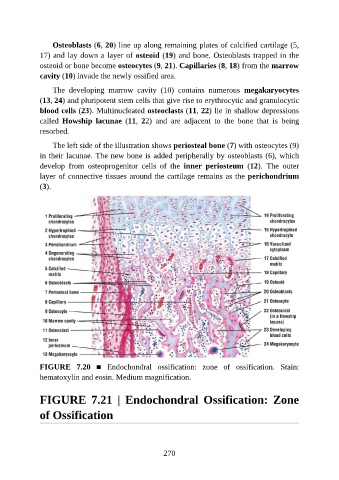
Osteoblasts (6, 20) line up along remaining plates of calcified cartilage (5,

               17) and lay down a layer of osteoid (19) and bone. Osteoblasts trapped in the
               osteoid or bone become osteocytes (9, 21). Capillaries (8, 18) from the marrow
               cavity (10) invade the newly ossified area.

                   The  developing  marrow  cavity  (10)  contains  numerous  megakaryocytes
               (13, 24) and pluripotent stem cells that give rise to erythrocytic and granulocytic

               blood cells (23). Multinucleated osteoclasts (11, 22) lie in shallow depressions
               called  Howship  lacunae  (11,  22)  and  are  adjacent  to  the  bone  that  is  being
               resorbed.

                   The left side of the illustration shows periosteal bone (7) with osteocytes (9)

               in their lacunae. The new bone is added peripherally by osteoblasts (6), which
               develop  from  osteoprogenitor  cells  of  the  inner  periosteum  (12).  The  outer
               layer of connective tissues around the cartilage remains as the perichondrium
               (3).


               FIGURE  7.20  ■  Endochondral  ossification:  zone  of  ossification.  Stain:
               hematoxylin and eosin. Medium magnification.


               FIGURE  7.21  |  Endochondral  Ossification:  Zone

               of Ossification


                                                          270