
Page 273 - Atlas of Histology with Functional Correlations
P. 273
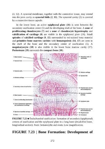
(4, 12). A synovial membrane, together with the connective tissue, may extend

               into the joint cavity as synovial folds (2, 13). The synovial cavity (3) is covered
               by a connective tissue capsule.

                   In  the  lower  bone,  an  active  epiphyseal  plate  (16)  is  seen  between  the
               secondary ossification center (5) and the developing shaft of the bone. A zone of
               proliferating chondrocytes (7) and a zone of chondrocyte hypertrophy and

               calcification  of  cartilage  (8)  are  visible  in  the  epiphyseal  plate  (16).  Small
               spicules of calcified cartilage (9, 15) surrounded by red-stained bony material
               and primitive bone marrow cavities with hematopoiesis (14, 17) are seen in
               the  shaft  of  the  bone  and  the  secondary  center  of  ossification  (5).  A

               megakaryocyte  (18)  is  also  visible  in  the  lower  bone  marrow  cavity  (17).
               Periosteum (19) surrounds the compact bone (10).


               FIGURE 7.22 ■ Endochondral ossification: formation of secondary (epiphyseal)
               centers of ossification and the epiphyseal plate in a long bone (decalcified bone,

               longitudinal section). Stain: hematoxylin and eosin. Low magnification.


               FIGURE 7.23 | Bone Formation: Development of


                                                          272