Page 278 - Atlas of Histology with Functional Correlations
P. 278
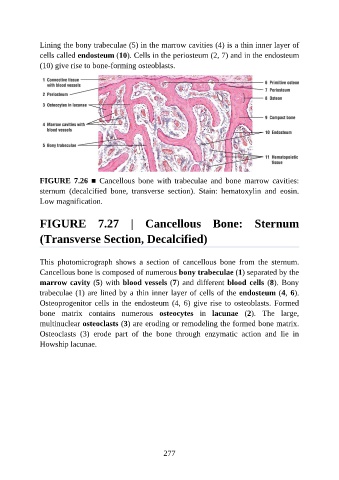
Lining the bony trabeculae (5) in the marrow cavities (4) is a thin inner layer of

               cells called endosteum (10). Cells in the periosteum (2, 7) and in the endosteum
               (10) give rise to bone-forming osteoblasts.


               FIGURE  7.26  ■  Cancellous  bone  with  trabeculae  and  bone  marrow  cavities:
               sternum  (decalcified  bone,  transverse  section).  Stain:  hematoxylin  and  eosin.

               Low magnification.


               FIGURE  7.27  |  Cancellous  Bone:  Sternum


               (Transverse Section, Decalcified)


               This  photomicrograph  shows  a  section  of  cancellous  bone  from  the  sternum.
               Cancellous bone is composed of numerous bony trabeculae (1) separated by the
               marrow cavity (5) with blood vessels (7) and different blood cells (8). Bony

               trabeculae (1) are lined by a thin inner layer of cells of the endosteum (4, 6).
               Osteoprogenitor cells in the endosteum (4, 6) give rise to osteoblasts. Formed
               bone  matrix  contains  numerous  osteocytes  in  lacunae  (2).  The  large,
               multinuclear osteoclasts (3) are eroding or remodeling the formed bone matrix.
               Osteoclasts  (3)  erode  part  of  the  bone  through  enzymatic  action  and  lie  in

               Howship lacunae.


                                                          277

