
Page 281 - Atlas of Histology with Functional Correlations
P. 281
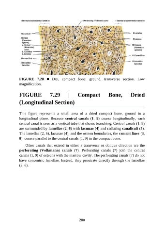
FIGURE  7.28  ■  Dry,  compact  bone:  ground,  transverse  section.  Low
               magnification.


               FIGURE                7.29          |     Compact                Bone,           Dried

               (Longitudinal Section)


               This  figure  represents  a  small  area  of  a  dried  compact  bone,  ground  in  a
               longitudinal  plane.  Because  central  canals  (1,  9)  course  longitudinally,  each
               central canal is seen as a vertical tube that shows branching. Central canals (1, 9)
               are surrounded by lamellae (2, 6) with lacunae (4) and radiating canaliculi (5).

               The lamellae (2, 6), lacunae (4), and the osteon boundaries, the cement lines (3,
               8), course parallel to the central canals (1, 9) in the compact bone.

                   Other canals that extend in either a transverse or oblique direction are the
               perforating  (Volkmann)  canals  (7).  Perforating  canals  (7)  join  the  central

               canals (1, 9) of osteons with the marrow cavity. The perforating canals (7) do not
               have concentric lamellae. Instead, they penetrate directly through the lamellae
               (2, 6).


                                                          280