
Page 282 - Atlas of Histology with Functional Correlations
P. 282
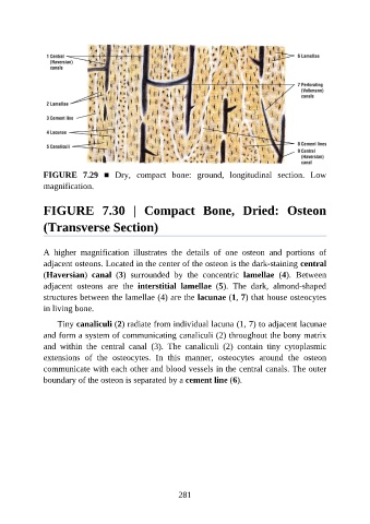
FIGURE  7.29  ■  Dry,  compact  bone:  ground,  longitudinal  section.  Low

               magnification.


               FIGURE  7.30  |  Compact  Bone,  Dried:  Osteon


               (Transverse Section)


               A  higher  magnification  illustrates  the  details  of  one  osteon  and  portions  of
               adjacent osteons. Located in the center of the osteon is the dark-staining central
               (Haversian)  canal  (3)  surrounded  by  the  concentric  lamellae  (4).  Between

               adjacent  osteons  are  the  interstitial  lamellae  (5).  The  dark,  almond-shaped
               structures between the lamellae (4) are the lacunae (1, 7) that house osteocytes
               in living bone.

                   Tiny canaliculi (2) radiate from individual lacuna (1, 7) to adjacent lacunae

               and form a system of communicating canaliculi (2) throughout the bony matrix
               and  within  the  central  canal  (3).  The  canaliculi  (2)  contain  tiny  cytoplasmic
               extensions  of  the  osteocytes.  In  this  manner,  osteocytes  around  the  osteon
               communicate with each other and blood vessels in the central canals. The outer

               boundary of the osteon is separated by a cement line (6).


                                                          281