
Page 341 - Atlas of Histology with Functional Correlations
P. 341
correspond to the structures illustrated in the cervical cord region in Figure 9.5.
These are the posterior median sulcus (15), anterior median fissure (22),
fasciculus gracilis (16) and fasciculus cuneatus (17) (seen in the mid-to-upper-
thoracic region of the spinal cord) of the posterior white column (16, 17),
lateral white column (7), central canal (9), and the gray commissure (18).
Associated with the posterior gray horns (6) are axons of the posterior roots (5),
and leaving the anterior gray horns (10, 20) are the axons (11, 21) of the
anterior roots (11).
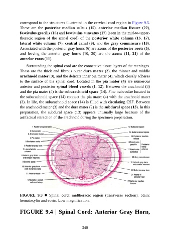
Surrounding the spinal cord are the connective tissue layers of the meninges.
These are the thick and fibrous outer dura mater (2), the thinner and middle
arachnoid mater (3), and the delicate inner pia mater (4), which closely adheres
to the surface of the spinal cord. Located in the pia mater (4) are numerous
anterior and posterior spinal blood vessels (1, 12). Between the arachnoid (3)
and the pia mater (4) is the subarachnoid space (14). Fine trabeculae located in
the subarachnoid space (14) connect the pia mater (4) with the arachnoid mater
(3). In life, the subarachnoid space (14) is filled with circulating CSF. Between
the arachnoid mater (3) and the dura mater (2) is the subdural space (13). In this
preparation, the subdural space (13) appears unusually large because of the
artifactual retraction of the arachnoid during the specimen preparation.
FIGURE 9.3 ■ Spinal cord: midthoracic region (transverse section). Stain:
hematoxylin and eosin. Low magnification.
FIGURE 9.4 | Spinal Cord: Anterior Gray Horn,
340