
Page 343 - Atlas of Histology with Functional Correlations
P. 343
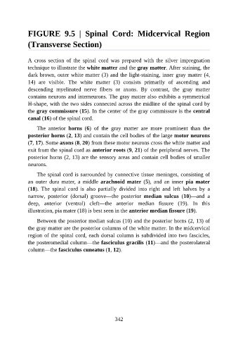
FIGURE  9.5  |  Spinal  Cord:  Midcervical  Region

               (Transverse Section)


               A  cross  section  of  the  spinal  cord  was  prepared  with  the  silver  impregnation
               technique to illustrate the white matter and the gray matter. After staining, the

               dark brown, outer white matter (3) and the light-staining, inner gray matter (4,
               14)  are  visible.  The  white  matter  (3)  consists  primarily  of  ascending  and
               descending  myelinated  nerve  fibers  or  axons.  By  contrast,  the  gray  matter
               contains neurons and interneurons. The gray matter also exhibits a symmetrical

               H-shape, with the two sides connected across the midline of the spinal cord by
               the gray commissure (15). In the center of the gray commissure is the central
               canal (16) of the spinal cord.

                   The  anterior  horns  (6)  of  the  gray  matter  are  more  prominent  than  the

               posterior horns (2, 13) and contain the cell bodies of the large motor neurons
               (7, 17). Some axons (8, 20) from these motor neurons cross the white matter and
               exit from the spinal cord as anterior roots (9, 21) of the peripheral nerves. The
               posterior horns (2, 13) are the sensory areas and contain cell bodies of smaller
               neurons.


                   The spinal cord is surrounded by connective tissue meninges, consisting of
               an  outer  dura  mater,  a  middle  arachnoid mater (5),  and  an  inner  pia  mater
               (18).  The  spinal  cord  is  also  partially  divided  into  right  and  left  halves  by  a
               narrow,  posterior  (dorsal)  groove—the  posterior  median  sulcus  (10)—and  a

               deep,  anterior  (ventral)  cleft—the  anterior  median  fissure  (19).  In  this
               illustration, pia mater (18) is best seen in the anterior median fissure (19).

                   Between the posterior median sulcus (10) and the posterior horns (2, 13) of
               the gray matter are the posterior columns of the white matter. In the midcervical

               region of the spinal cord, each dorsal column is subdivided into two fascicles,
               the posteromedial column—the fasciculus gracilis (11)—and the posterolateral
               column—the fasciculus cuneatus (1, 12).


                                                          342