
Page 342 - Atlas of Histology with Functional Correlations
P. 342
Motor Neurons, and Adjacent Anterior White
Matter
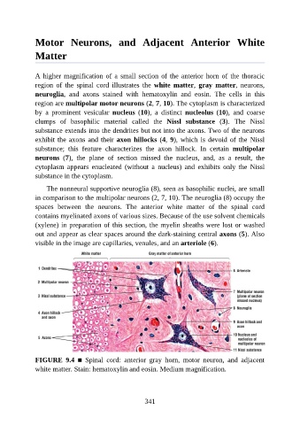
A higher magnification of a small section of the anterior horn of the thoracic
region of the spinal cord illustrates the white matter, gray matter, neurons,
neuroglia, and axons stained with hematoxylin and eosin. The cells in this
region are multipolar motor neurons (2, 7, 10). The cytoplasm is characterized
by a prominent vesicular nucleus (10), a distinct nucleolus (10), and coarse
clumps of basophilic material called the Nissl substance (3). The Nissl
substance extends into the dendrites but not into the axons. Two of the neurons
exhibit the axons and their axon hillocks (4, 9), which is devoid of the Nissl
substance; this feature characterizes the axon hillock. In certain multipolar
neurons (7), the plane of section missed the nucleus, and, as a result, the
cytoplasm appears enucleated (without a nucleus) and exhibits only the Nissl
substance in the cytoplasm.
The nonneural supportive neuroglia (8), seen as basophilic nuclei, are small
in comparison to the multipolar neurons (2, 7, 10). The neuroglia (8) occupy the
spaces between the neurons. The anterior white matter of the spinal cord
contains myelinated axons of various sizes. Because of the use solvent chemicals
(xylene) in preparation of this section, the myelin sheaths were lost or washed
out and appear as clear spaces around the dark-staining central axons (5). Also
visible in the image are capillaries, venules, and an arteriole (6).
FIGURE 9.4 ■ Spinal cord: anterior gray horn, motor neuron, and adjacent
white matter. Stain: hematoxylin and eosin. Medium magnification.
341