
Page 346 - Atlas of Histology with Functional Correlations
P. 346
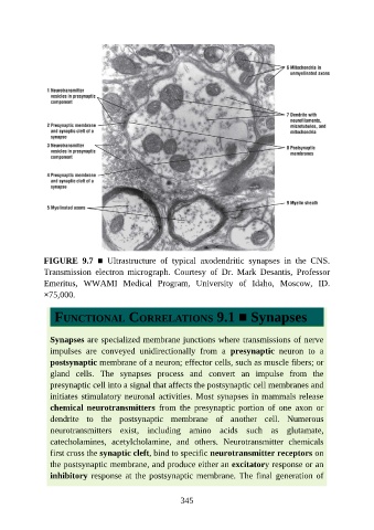
FIGURE  9.7  ■  Ultrastructure  of  typical  axodendritic  synapses  in  the  CNS.
               Transmission  electron  micrograph.  Courtesy  of  Dr.  Mark  Desantis,  Professor

               Emeritus,  WWAMI  Medical  Program,  University  of  Idaho,  Moscow,  ID.
               ×75,000.


                  FUNCTIONAL CORRELATIONS 9.1 ■ Synapses


                 Synapses are specialized membrane junctions where transmissions of nerve
                 impulses  are  conveyed  unidirectionally  from  a  presynaptic  neuron  to  a
                 postsynaptic membrane of a neuron; effector cells, such as muscle fibers; or

                 gland  cells.  The  synapses  process  and  convert  an  impulse  from  the
                 presynaptic cell into a signal that affects the postsynaptic cell membranes and
                 initiates stimulatory neuronal activities. Most synapses in mammals release
                 chemical neurotransmitters  from  the  presynaptic  portion  of  one  axon  or
                 dendrite  to  the  postsynaptic  membrane  of  another  cell.  Numerous

                 neurotransmitters  exist,  including  amino  acids  such  as  glutamate,
                 catecholamines,  acetylcholamine,  and  others.  Neurotransmitter  chemicals
                 first cross the synaptic cleft, bind to specific neurotransmitter receptors on

                 the postsynaptic membrane, and produce either an excitatory response or an
                 inhibitory response at the postsynaptic membrane. The final generation of


                                                          345