
Page 351 - Atlas of Histology with Functional Correlations
P. 351
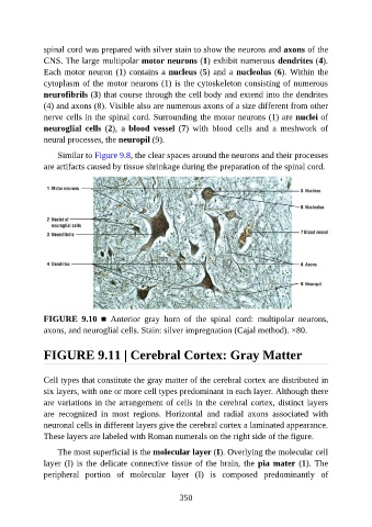
spinal cord was prepared with silver stain to show the neurons and axons of the

               CNS. The large multipolar motor neurons (1) exhibit numerous dendrites (4).
               Each motor neuron (1) contains a nucleus (5) and a nucleolus (6). Within the
               cytoplasm of the motor neurons (1) is the cytoskeleton consisting of numerous
               neurofibrils (3) that course through the cell body and extend into the dendrites
               (4) and axons (8). Visible also are numerous axons of a size different from other

               nerve cells in the spinal cord. Surrounding the motor neurons (1) are nuclei of
               neuroglial  cells  (2),  a  blood  vessel  (7)  with  blood  cells  and  a  meshwork  of
               neural processes, the neuropil (9).

                   Similar to Figure 9.8, the clear spaces around the neurons and their processes

               are artifacts caused by tissue shrinkage during the preparation of the spinal cord.


               FIGURE  9.10  ■  Anterior  gray  horn  of  the  spinal  cord:  multipolar  neurons,
               axons, and neuroglial cells. Stain: silver impregnation (Cajal method). ×80.


               FIGURE 9.11 | Cerebral Cortex: Gray Matter


               Cell types that constitute the gray matter of the cerebral cortex are distributed in

               six layers, with one or more cell types predominant in each layer. Although there
               are variations in the arrangement of cells in the cerebral cortex, distinct layers
               are  recognized  in  most  regions.  Horizontal  and  radial  axons  associated  with
               neuronal cells in different layers give the cerebral cortex a laminated appearance.
               These layers are labeled with Roman numerals on the right side of the figure.


                   The most superficial is the molecular layer (I). Overlying the molecular cell
               layer (I) is the delicate connective tissue of the brain, the pia mater (1). The
               peripheral  portion  of  molecular  layer  (I)  is  composed  predominantly  of


                                                          350