
Page 356 - Atlas of Histology with Functional Correlations
P. 356
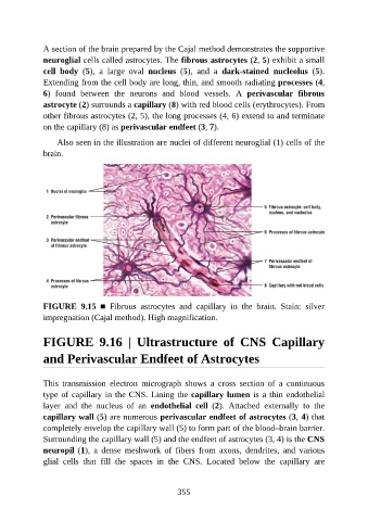
A section of the brain prepared by the Cajal method demonstrates the supportive

               neuroglial cells called astrocytes. The fibrous astrocytes (2, 5) exhibit a small
               cell  body  (5),  a  large  oval  nucleus  (5),  and  a  dark-stained  nucleolus  (5).
               Extending from the cell body are long, thin, and smooth radiating processes (4,
               6)  found  between  the  neurons  and  blood  vessels.  A  perivascular  fibrous
               astrocyte (2) surrounds a capillary (8) with red blood cells (erythrocytes). From

               other fibrous astrocytes (2, 5), the long processes (4, 6) extend to and terminate
               on the capillary (8) as perivascular endfeet (3, 7).

                   Also seen in the illustration are nuclei of different neuroglial (1) cells of the
               brain.


               FIGURE  9.15  ■  Fibrous  astrocytes  and  capillary  in  the  brain.  Stain:  silver

               impregnation (Cajal method). High magnification.


               FIGURE  9.16  |  Ultrastructure  of  CNS  Capillary


               and Perivascular Endfeet of Astrocytes


               This  transmission  electron  micrograph  shows  a  cross  section  of  a  continuous
               type of capillary in the CNS. Lining the capillary lumen is a thin endothelial
               layer  and  the  nucleus  of  an  endothelial  cell  (2).  Attached  externally  to  the

               capillary wall (5) are numerous perivascular endfeet of astrocytes (3, 4) that
               completely envelop the capillary wall (5) to form part of the blood–brain barrier.
               Surrounding the capillary wall (5) and the endfeet of astrocytes (3, 4) is the CNS
               neuropil (1),  a  dense  meshwork  of  fibers  from  axons,  dendrites,  and  various
               glial  cells  that  fill  the  spaces  in  the  CNS.  Located  below  the  capillary  are


                                                          355