
Page 360 - Atlas of Histology with Functional Correlations
P. 360
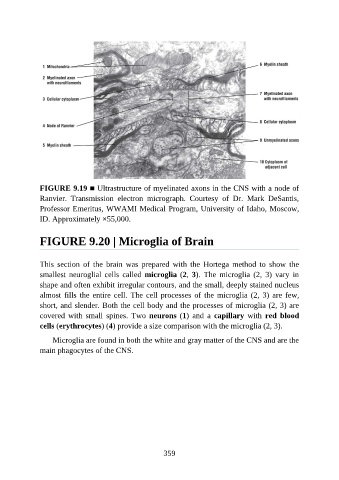
FIGURE 9.19 ■ Ultrastructure of myelinated axons in the CNS with a node of
               Ranvier.  Transmission  electron  micrograph.  Courtesy  of  Dr.  Mark  DeSantis,
               Professor Emeritus, WWAMI Medical Program, University of Idaho, Moscow,
               ID. Approximately ×55,000.


               FIGURE 9.20 | Microglia of Brain


               This  section  of  the  brain  was  prepared  with  the  Hortega  method  to  show  the

               smallest  neuroglial  cells  called  microglia (2, 3).  The  microglia  (2,  3)  vary  in
               shape and often exhibit irregular contours, and the small, deeply stained nucleus
               almost fills the entire cell. The cell processes of the microglia (2, 3) are few,
               short, and slender. Both the cell body and the processes of microglia (2, 3) are
               covered  with  small  spines.  Two  neurons (1)  and  a  capillary  with  red  blood

               cells (erythrocytes) (4) provide a size comparison with the microglia (2, 3).

                   Microglia are found in both the white and gray matter of the CNS and are the
               main phagocytes of the CNS.


                                                          359