
Page 357 - Atlas of Histology with Functional Correlations
P. 357
myelinated axons (6) that were myelinated in the CNS by oligodendrocytes (not
illustrated).
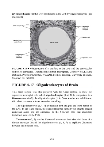
FIGURE 9.16 ■ Ultrastructure of a capillary in the CNS and the perivascular
endfeet of astrocytes. Transmission electron micrograph. Courtesy of Dr. Mark
DeSantis, Professor Emeritus, WWAMI. Medical Program, University of Idaho,
Moscow, ID. ×20,000.
FIGURE 9.17 | Oligodendrocytes of Brain
This brain section was also prepared with the Cajal method to show the
supportive neuroglial cells called oligodendrocytes (1, 4, 7). In comparison to a
fibrous astrocyte (3), the oligodendrocytes (1, 4, 7) are smaller and exhibit few,
thin, short processes without excessive branching.
The oligodendrocytes (1, 4, 7) are found in both the gray and white matter of
the CNS. In the white matter, the oligodendrocytes form myelin sheaths around
numerous axons and are analogous to the Schwann cells that myelinate
individual axons in the PNS.
Two neurons (2, 6) are also illustrated to contrast their size with those of a
fibrous astrocyte (3) and the oligodendrocytes (1, 4, 7). A capillary (5) passes
between the different cells.
356