
Page 353 - Atlas of Histology with Functional Correlations
P. 353
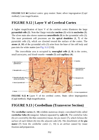
FIGURE 9.11 ■ Cerebral cortex: gray matter: Stain: silver impregnation (Cajal

               method). Low magnification.


               FIGURE 9.12 | Layer V of Cerebral Cortex


               A  higher  magnification  of  layer  V  of  the  cerebral  cortex  illustrates  the  large

               pyramidal cells (3). Note the l large vesicular nucleus (3) with its nucleolus (3).
               The silver stain also shows numerous neurofibrils (9) in the pyramidal cells (3).
               The  most  prominent  cell  processes  are  the  apical  dendrites  (1,  7)  of  the
               pyramidal  cells  (3),  which  are  directed  toward  the  surface  of  the  cortex.  The

               axons (4, 10) of the pyramidal cells (3) arise from the base of the cell body and
               pass into the white matter (see Fig. 9.11 [10]).

                   The intercellular area is occupied by neuroglial cells (2, 8)  in  the  cortex,
               small astrocytes, and blood vessels—venule (5) and capillary (6).


               FIGURE  9.12  ■  Layer  V  of  the  cerebral  cortex.  Stain:  silver  impregnation

               (Cajal method). High magnification.


               FIGURE 9.13 | Cerebellum (Transverse Section)


               The cerebellar cortex (1, 10) exhibits numerous deeply convoluted folds called
               cerebellar folia (6) (singular: folium) separated by sulci (9). The cerebellar folia
               (6) are covered by the thin connective tissue, the pia mater (7), which follows the
               surface of each folium (6) into the adjacent sulci (9). The detachment of the pia
               mater  (7)  from  the  cerebellar  cortex  (1,  10)  is  an  artifact  caused  by  tissue

               fixation and preparation.


                                                          352