
Page 350 - Atlas of Histology with Functional Correlations
P. 350
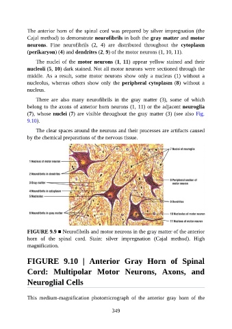
The anterior horn of the spinal cord was prepared by silver impregnation (the

               Cajal method) to demonstrate neurofibrils in both the gray matter and motor
               neurons.  Fine  neurofibrils  (2,  4)  are  distributed  throughout  the  cytoplasm
               (perikaryon) (4) and dendrites (2, 9) of the motor neurons (1, 10, 11).

                   The  nuclei  of  the  motor neurons (1, 11)  appear  yellow  stained  and  their
               nucleoli (5, 10) dark stained. Not all motor neurons were sectioned through the

               middle.  As  a  result,  some  motor  neurons  show  only  a  nucleus  (1)  without  a
               nucleolus,  whereas  others  show  only  the  peripheral cytoplasm (8)  without  a
               nucleus.

                   There  are  also  many  neurofibrils  in  the  gray  matter  (3),  some  of  which

               belong to the axons of anterior horn neurons (1, 11) or the adjacent neuroglia
               (7), whose nuclei (7) are visible throughout the gray matter (3) (see also Fig.
               9.10).

                   The clear spaces around the neurons and their processes are artifacts caused
               by the chemical preparations of the nervous tissue.


               FIGURE 9.9 ■ Neurofibrils and motor neurons in the gray matter of the anterior

               horn  of  the  spinal  cord.  Stain:  silver  impregnation  (Cajal  method).  High
               magnification.


               FIGURE  9.10  |  Anterior  Gray  Horn  of  Spinal

               Cord:  Multipolar  Motor  Neurons,  Axons,  and

               Neuroglial Cells


               This  medium-magnification  photomicrograph  of  the  anterior  gray  horn  of  the


                                                          349