
Page 354 - Atlas of Histology with Functional Correlations
P. 354
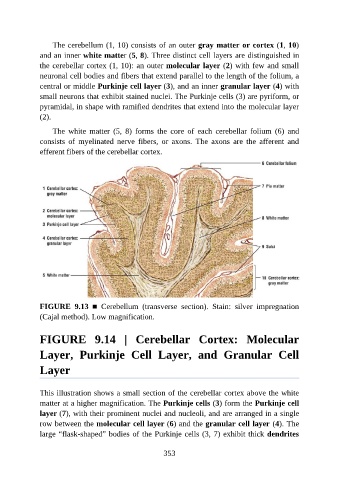
The cerebellum (1, 10) consists of an outer gray matter or cortex (1, 10)

               and an inner white matter (5, 8). Three distinct cell layers are distinguished in
               the cerebellar cortex (1, 10): an outer molecular layer (2) with few and small
               neuronal cell bodies and fibers that extend parallel to the length of the folium, a
               central or middle Purkinje cell layer (3), and an inner granular layer (4) with
               small neurons that exhibit stained nuclei. The Purkinje cells (3) are pyriform, or

               pyramidal, in shape with ramified dendrites that extend into the molecular layer
               (2).

                   The  white  matter  (5,  8)  forms  the  core  of  each  cerebellar  folium  (6)  and
               consists  of  myelinated  nerve  fibers,  or  axons.  The  axons  are  the  afferent  and

               efferent fibers of the cerebellar cortex.


               FIGURE  9.13  ■  Cerebellum  (transverse  section).  Stain:  silver  impregnation

               (Cajal method). Low magnification.


               FIGURE  9.14  |  Cerebellar  Cortex:  Molecular

               Layer,  Purkinje  Cell  Layer,  and  Granular  Cell

               Layer


               This illustration shows a small section of the cerebellar cortex above the white
               matter at a higher magnification. The Purkinje cells (3) form the Purkinje cell
               layer (7), with their prominent nuclei and nucleoli, and are arranged in a single
               row between the molecular cell layer (6) and the granular cell layer (4). The
               large “flask-shaped” bodies of the Purkinje cells (3, 7) exhibit thick dendrites


                                                          353