
Page 358 - Atlas of Histology with Functional Correlations
P. 358
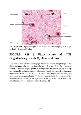
FIGURE 9.17 ■ Oligodendrocytes of the brain. Stain: silver impregnation (Cajal
               method). High magnification.


               FIGURE                9.18         |      Ultrastructure                 of        CNS

               Oligodendrocyte with Myelinated Axons


               This  transmission  electron  micrograph  illustrates  internal  morphology  of  the
               oligodendrocyte  (2),  the  myelin-producing  cell  of  the  CNS.  The  cytoplasm
               exhibits  a  well-developed  granular  endoplasmic  reticulum  (3,  5),  a  Golgi
               apparatus (6), and numerous free ribosomes around the organelles. Numerous

               myelinated  axons  (1,  4,  8),  cut  in  cross  and  longitudinal  sections,  are
               surrounded with myelin sheaths (7) that are associated with the cytoplasm of the
               oligodendrocyte. Located in the myelinated axons (1, 4) are oval, dark-staining
               mitochondria (4) and numerous neurofilaments (8).


                                                          357