
Page 359 - Atlas of Histology with Functional Correlations
P. 359
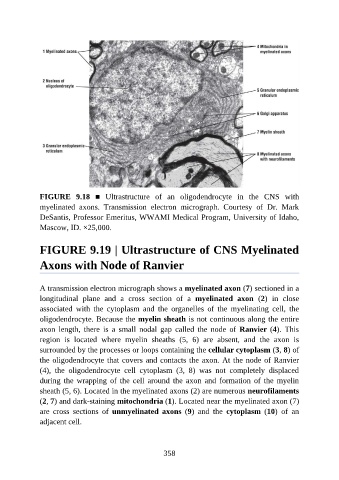
FIGURE  9.18  ■  Ultrastructure  of  an  oligodendrocyte  in  the  CNS  with

               myelinated  axons.  Transmission  electron  micrograph.  Courtesy  of  Dr.  Mark
               DeSantis, Professor Emeritus, WWAMI Medical Program, University of Idaho,
               Mascow, ID. ×25,000.


               FIGURE 9.19 | Ultrastructure of CNS Myelinated

               Axons with Node of Ranvier


               A transmission electron micrograph shows a myelinated axon (7) sectioned in a

               longitudinal  plane  and  a  cross  section  of  a  myelinated  axon  (2)  in  close
               associated  with  the  cytoplasm  and  the  organelles  of  the  myelinating  cell,  the
               oligodendrocyte. Because the myelin sheath is not continuous along the entire

               axon  length,  there  is  a  small  nodal  gap  called  the  node  of  Ranvier (4).  This
               region  is  located  where  myelin  sheaths  (5,  6)  are  absent,  and  the  axon  is
               surrounded by the processes or loops containing the cellular cytoplasm (3, 8) of
               the oligodendrocyte that covers and contacts the axon. At the node of Ranvier
               (4),  the  oligodendrocyte  cell  cytoplasm  (3,  8)  was  not  completely  displaced

               during  the  wrapping  of  the  cell  around  the  axon  and  formation  of  the  myelin
               sheath (5, 6). Located in the myelinated axons (2) are numerous neurofilaments
               (2, 7) and dark-staining mitochondria (1). Located near the myelinated axon (7)

               are  cross  sections  of  unmyelinated  axons  (9)  and  the  cytoplasm  (10)  of  an
               adjacent cell.


                                                          358