
Page 361 - Atlas of Histology with Functional Correlations
P. 361
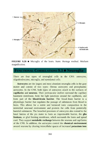
FIGURE  9.20  ■  Microglia  of  the  brain.  Stain:  Hortega  method.  Medium
               magnification.


                  FUNCTIONAL CORRELATIONS 9.3 ■ Neuroglia


                 There  are  four  types  of  neuroglial  cells  in  the  CNS:  astrocytes,
                 oligodendrocytes, microglia, and ependymal cells.

                     Astrocytes are the largest and most abundant neuroglia cells in the gray

                 matter  and  consist  of  two  types:  fibrous  astrocytes  and  protoplasmic
                 astrocytes.  In  the  CNS,  both  types  of  astrocytes  attach  to  the  surfaces  of
                 capillaries  and  neurons.  Their  perivascular  endfeet  surround  the  capillary
                 basement  membrane,  form  the  tight  junctions  around  the  capillaries,  and

                 form  part  of  the  blood–brain  barrier.  The  blood–brain  barrier  is  a
                 physiologic  barrier  that  regulates  the  passage  of  substances  from  blood  to
                 brain.  This  allows  for  a  stable  and  balanced  ionic  composition  in  the
                 interstitial  neuronal  environment  and  protects  the  cells  from  potentially

                 harmful substances. The branched processes of astrocytes also extend to the
                 basal  lamina  of  the  pia  mater  to  form  an  impermeable  barrier,  the  glia
                 limitans, or glial limiting membrane, which surrounds the brain and spinal
                 cord. They support metabolic exchange between the neurons and capillaries

                 of  the  CNS.  In  addition,  the  astrocytes  control  the  chemical environment
                 around neurons by clearing intercellular spaces of increased potassium ions


                                                          360